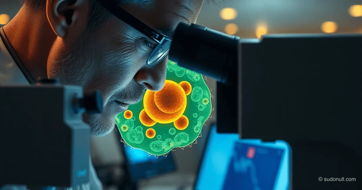
Domestic Laser Microscope with 0.1 nm Resolution Completes Testing for Cell Research
Laser microscope with resolution up to 0.1 nm vertically and 10–100 nm in the lateral plane has completed testing at the National Medical Research Center named after V. A. Almazov. The device allows visualization of the internal structure of living cells without damaging the plasma membrane. It is used for developing oncology drugs and controlling the topology of integrated circuits in microelectronics.
Technical Specifications and Advantages
Optical resolution of the microscope: 10–100 nm in the lateral plane, 0.1 nm vertically. This provides ultra-precise visualization of subcellular structures inaccessible to standard optical methods. Development is underway at the Ural Optical-Mechanical Plant named after E. S. Yalamov.
Comparison with counterparts:
- Swiss counterpart: higher image formation speed, but lower quality.
- American counterpart: high quality, but low operating speed.
The Russian microscope combines high resolution and acceptable performance, positioning it as the first device in its class in terms of overall characteristics.
Patent for the industrial design registered in 2015 by the Federal Service for Intellectual Property. Investments: 550 million rubles, including 375 million in subsidized budget funds.
Applications in Oncology and Diagnostics
The microscope is used for noninvasive analysis of healthy and tumor cells. Testing at the Almazov Center lasted about two years. The device is already being used in diagnosing cancer and cardiovascular diseases.
The process of testing anti-tumor drugs includes the following stages:
- Studying the morphology of cancer cells: shape, size, internal structure.
- Adding microdoses of the drug to the biomaterial.
- Analyzing the impact on malignant cells.
- Determining the minimum effective dose for single administration.
Five units have been produced so far. Four are undergoing testing at other institutions, with each subsequent one adapted to the customer's needs.
Prospects in Microelectronics and Export
In microelectronics, the microscope assesses the quality of IC topology—a key parameter for processor performance. 0.1 nm accuracy allows detecting defects at the nanoscale without destroying samples.
Export readiness: the company is accepting orders for manufacturing to the specifications of foreign customers. Valeria Tonkoshkurova, Development Director for Medical Projects at the Shvabe holding, confirmed its export potential.
Evgeny Shlyakhto, CEO of the Almazov Center, noted its effectiveness in real clinical scenarios.
Key Points
- 0.1 nm vertical resolution—a world achievement for noninvasive cellular visualization.
- Applications: oncology (drug testing), microelectronics (IC topology control).
- 5 units produced, serial manufacturing on order.
- Investments 550 million rubles, patent from 2015.
- Ready for export without additional modifications.
The technology opens new opportunities for precision medicine and nanotechnology, where subcellular resolution is required.
— Editorial Team










No comments yet.