Vision correction equipment
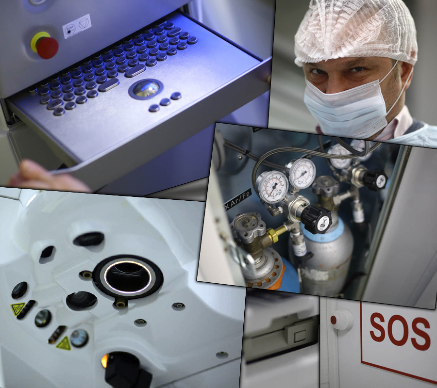
This is a photo excursion , 70 photographs from two operating units for laser vision correction will continue.
Different options for expander:
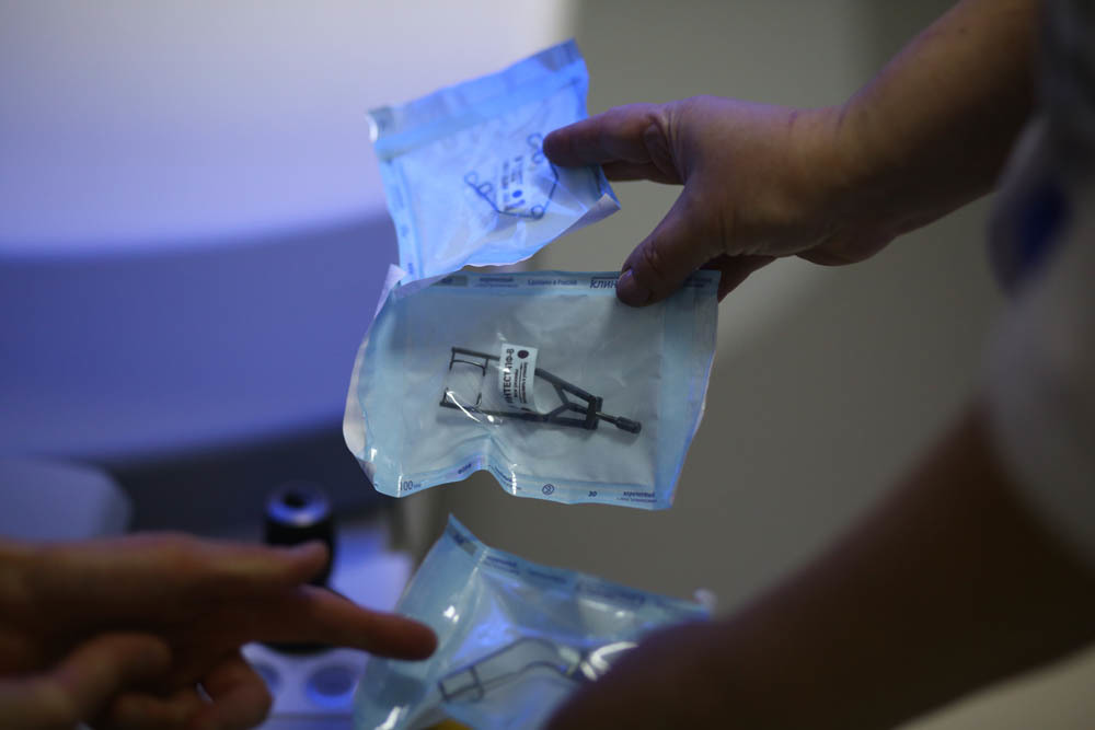

Packaging with a disposable blade, which is charged in a microkeratome to form a valve in the cornea during mechanical (not laser) operations:

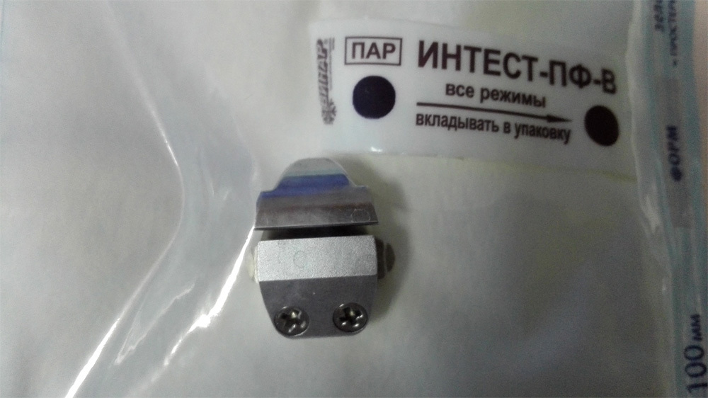
But the microkeratome head, where the blade is inserted:
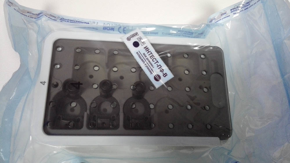
This is a set of "accessories" for the working part - it is reusable:

Motor for driving a head with a blade:

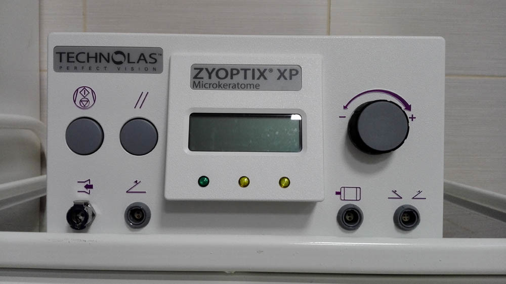
And this is a console with an indicator of operating parameters:
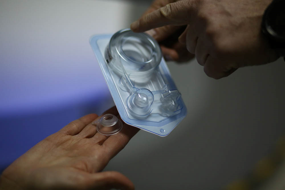
And now let's compare it with tools for SMILE. This is what disposable packaging with a cone looks like, in the same place - a vacuum tube for the removal of gas:
Spatula for separation on a scale:
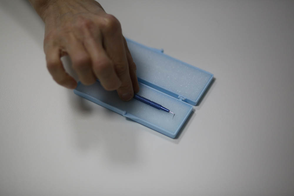
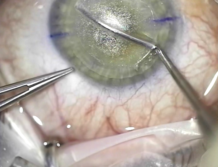
The very first spatula was made by Dr. Bloom, who invented technology with Walter Secundo. Today they do not use it, because it was spicy, because then they were cutting at other frequencies, there were no modern lasers. Now the cut is much cleaner, and there are already about 10 different modifications of tools. The most popular is according to Chansu (this is a Thai surgeon who performs a wild number of operations, 8 or 10 thousand one per year, he has a “conveyor” type clinic in Bangkok). There are interesting Russian ones, in particular, Kostin's spatula from Yekaterinburg. There is no huge difference between them. Walter enjoys just like in the photo, this is his author’s model just under his arm. Let me remind you that this is what it looks like (in the photo below during the operation, the Chansu spatula):
Microsponges of a hydrophilic material that someone rattled in the second post (not the blade):

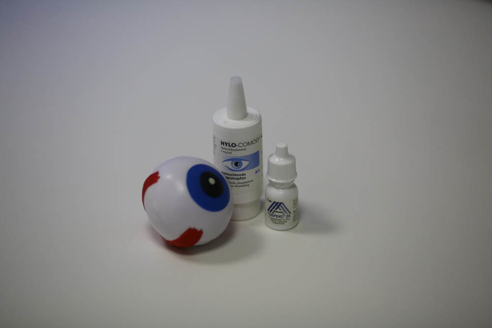
The fact that the patient takes home after correction:
Here is the disposable scalpel to the cornea, which is almost not needed (its functions are performed by the laser):

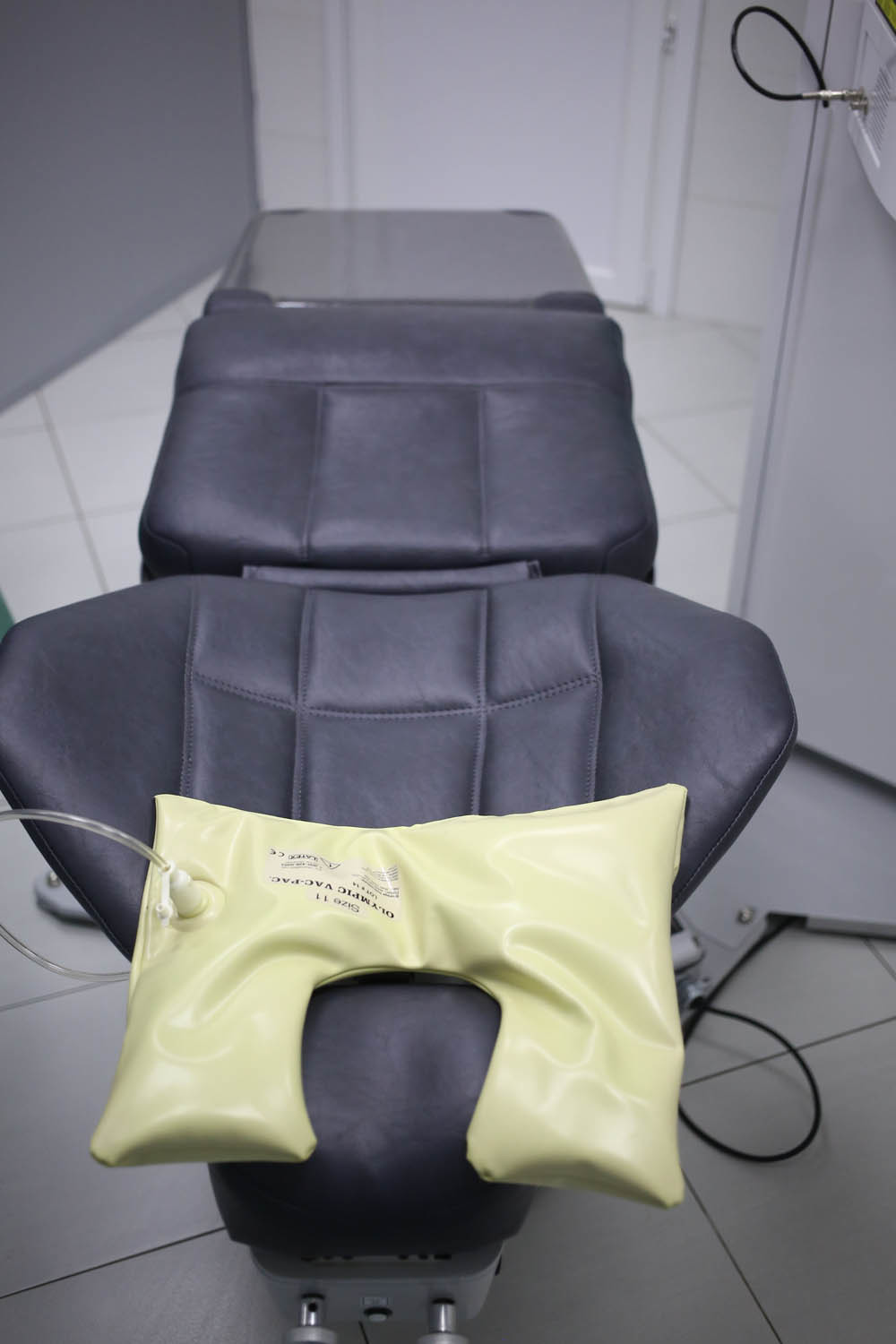
For the patient begins all after the corridor with such a cot. Here the patient lies a little before the correction (to calm the motility) and rests a couple of hours after.
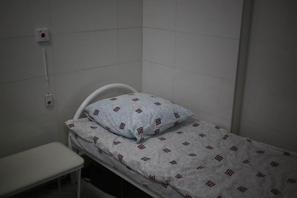
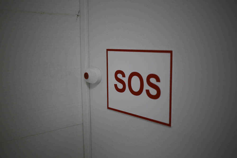
On the wall are two buttons: call sister and SOS. SOS includes an alarm in all rooms, except for other operating rooms.
On the wall pictures for staring:
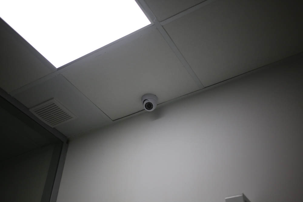
There is a video camera monitoring the patient's condition in the ward. The image is broadcast on the post of a nurse, so that we always know what is happening with the patient. It is especially needed for those long operations, after which the patient wakes up after anesthesia is not quite in a normal state of consciousness.
The first operating with excimer laser:
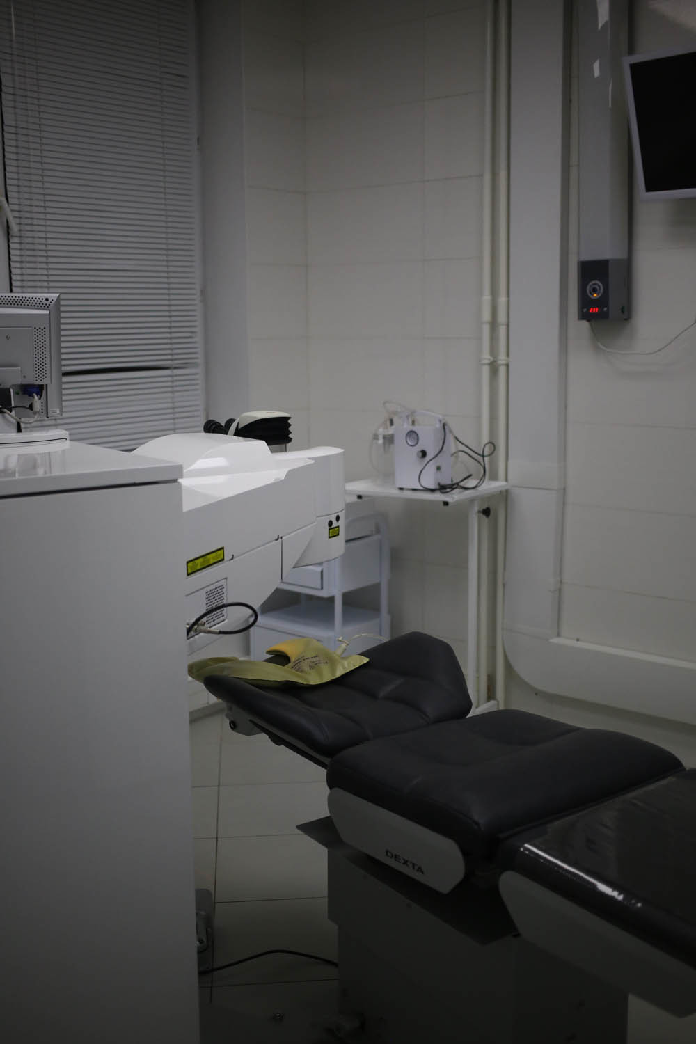
Couch (more precisely, the operating table, it - the smart bed):
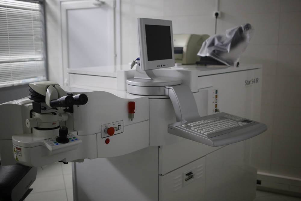
Excimer laser:

It is the "face" to the patient (this part is placed under a microscope):
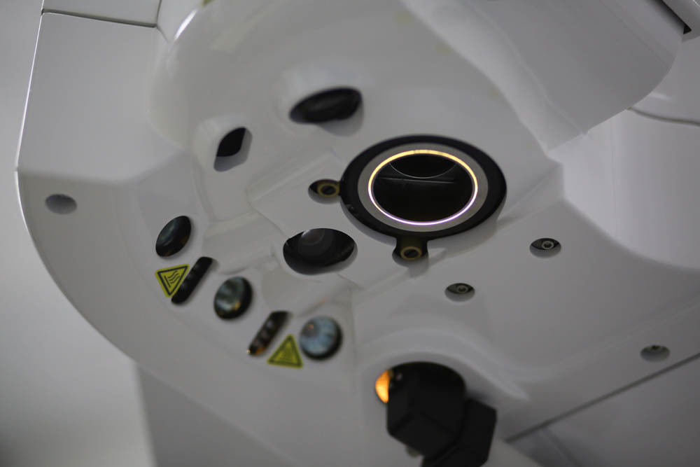
This is how the laser is visible to the surgeon:
In this microscope is conducted surveillance . The data is also displayed for the assistant on the screen on the operating room wall.
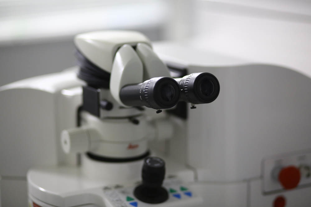
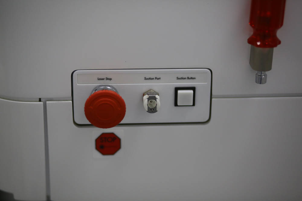
On hand at the doctor - laser stop button:
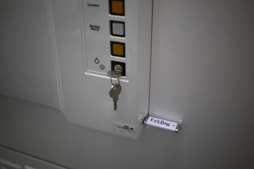
This is the key to start and pre-calculated profile for the operation:
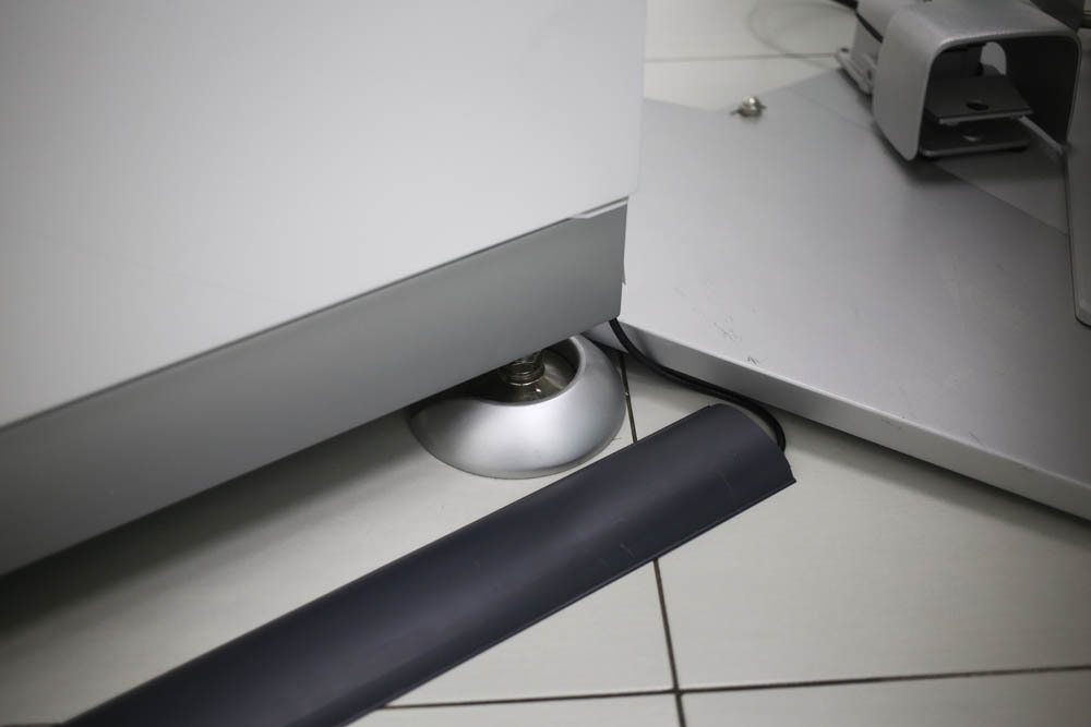
Vibration protection:
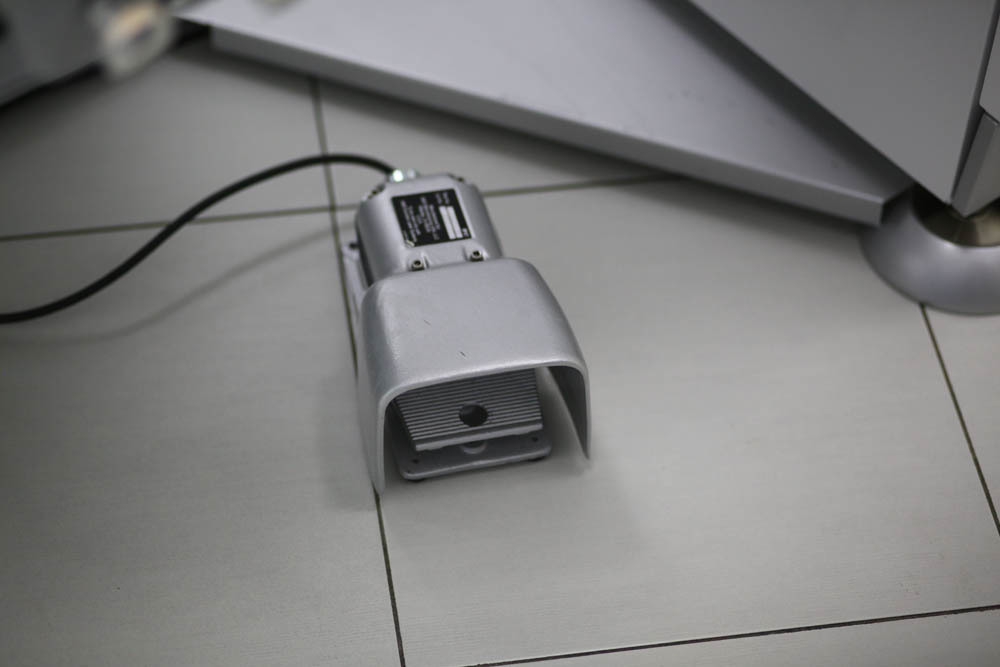
Laser control pedal (during the operation it is under the doctor's foot):
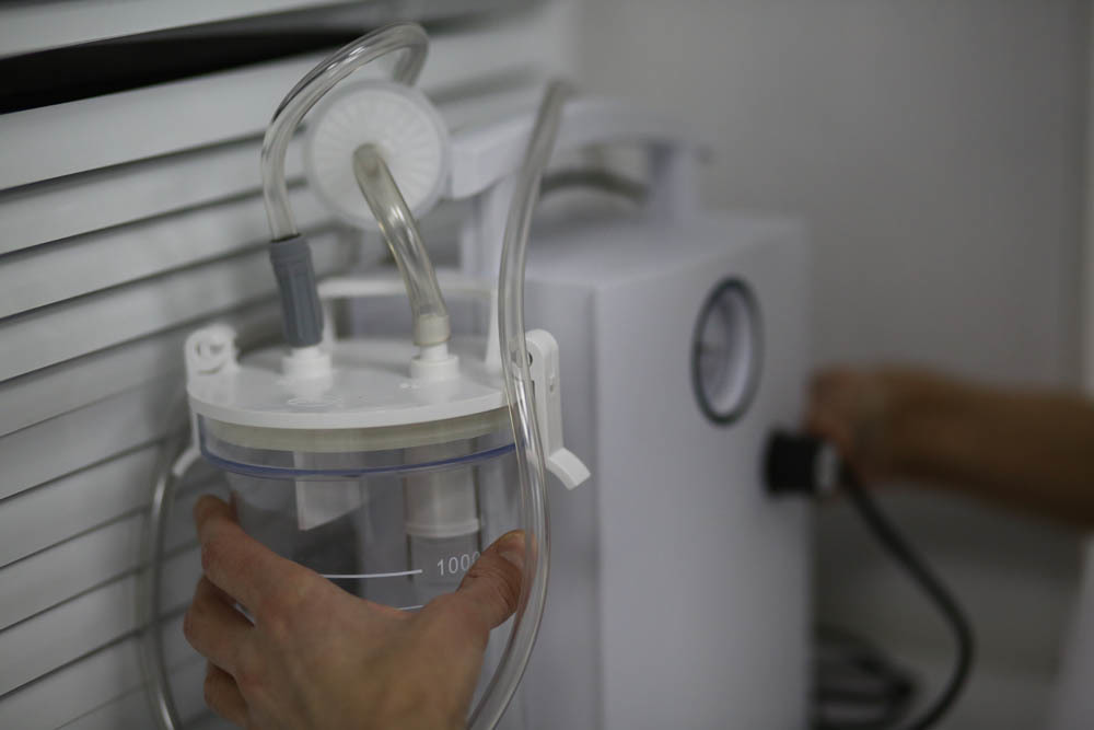
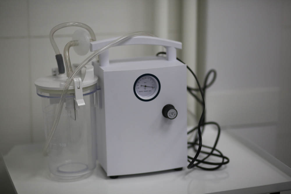
Vacuum suction to remove excess moisture and fatty secretions of the glands:
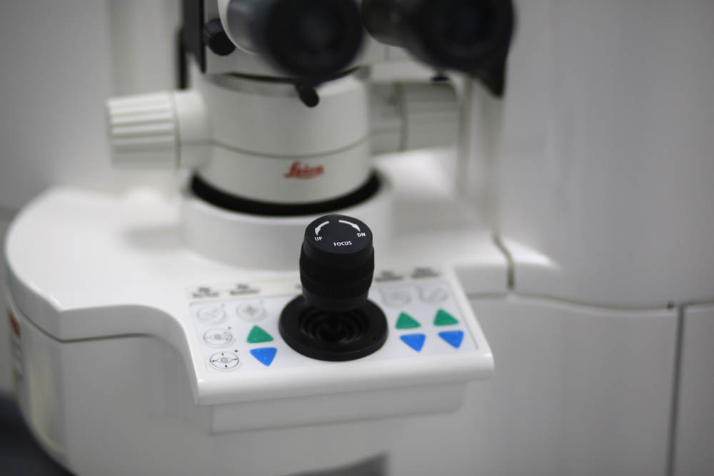
Excimer laser control
knob : Laser calibration meter (laser burns special test calibration lenses of various dioptries).
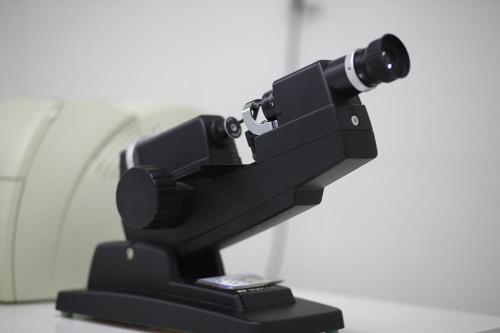
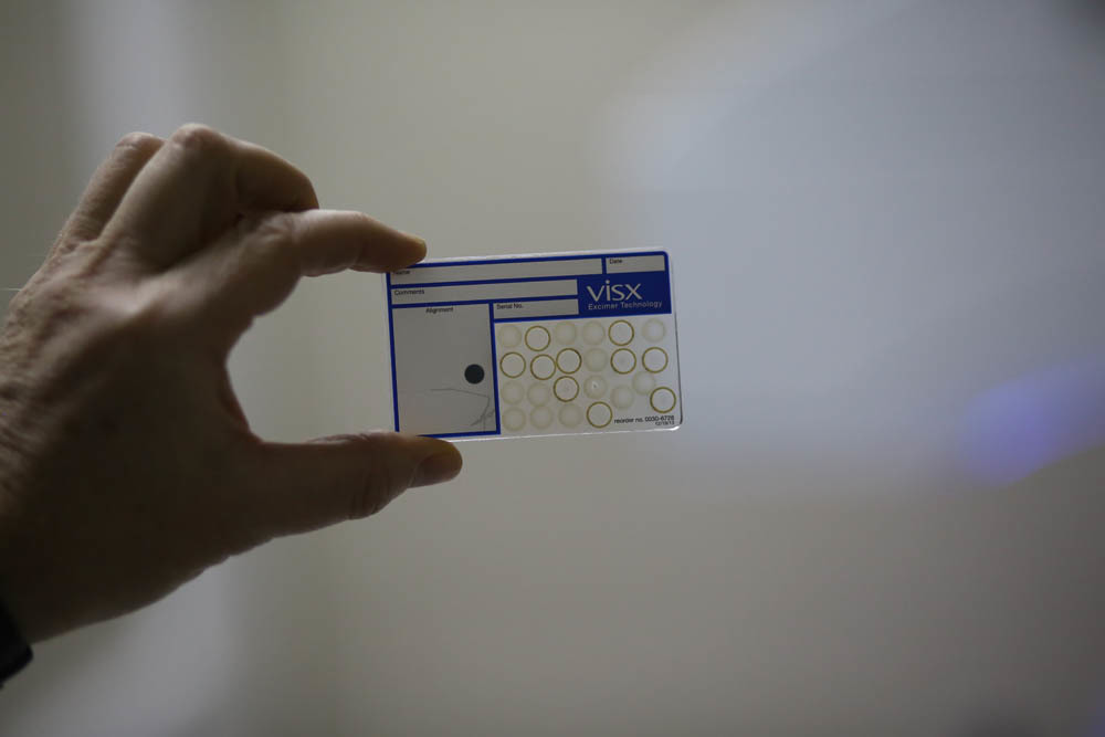
These are the cards, the laser burns the lenses of different dioptries on them:
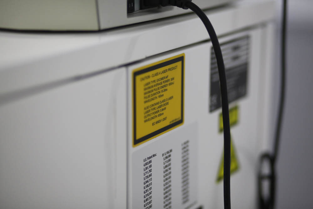
Warned warnings:
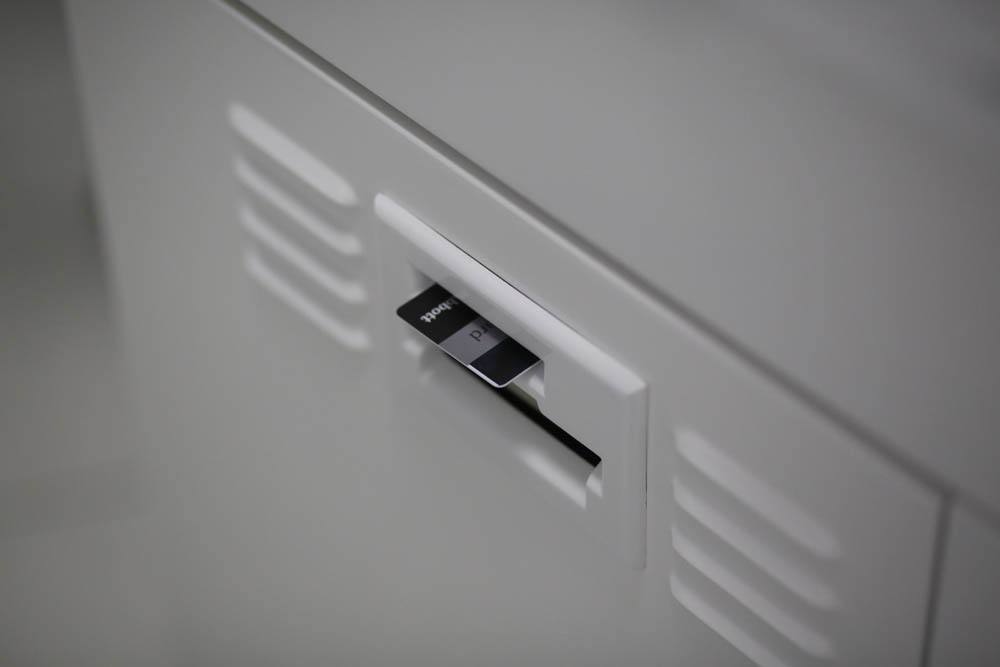
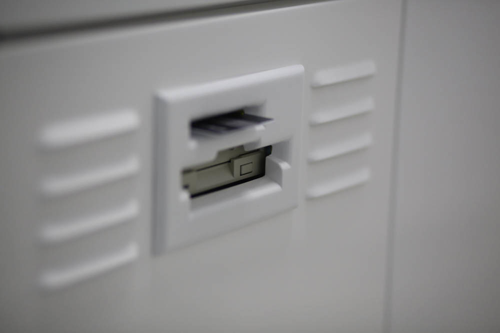
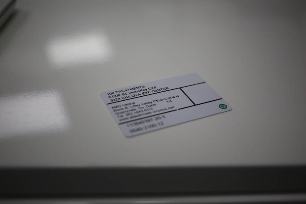
A license card (the femtosecond laser has a fully digital license):
Guess who is under it: The
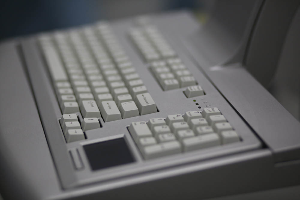
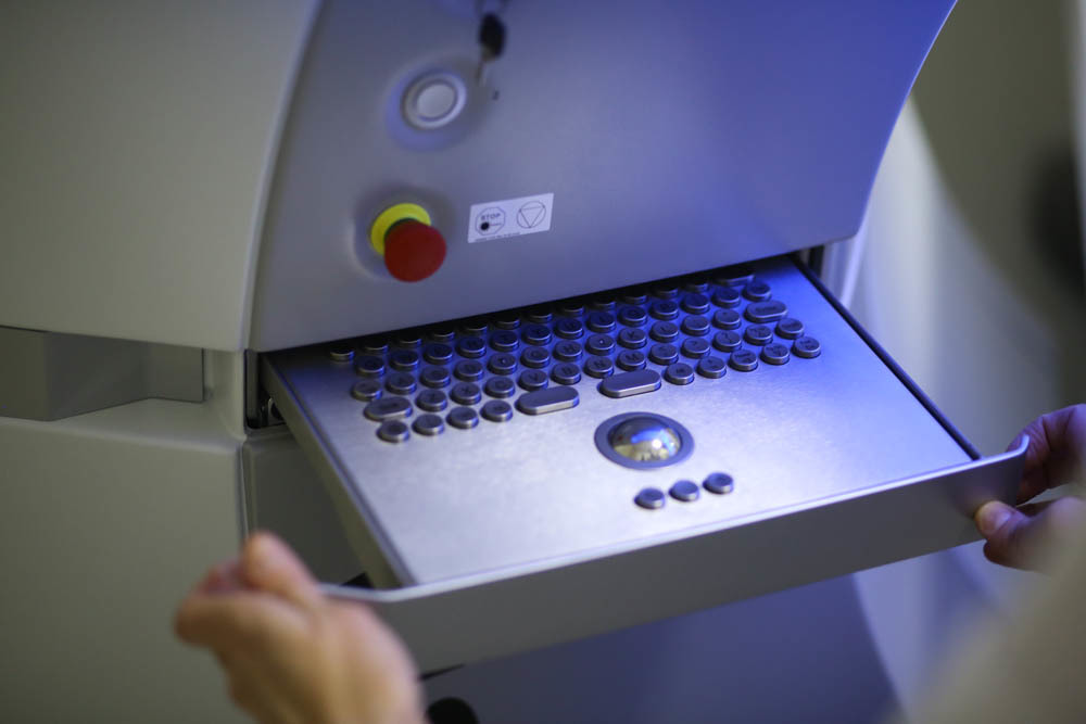
keyboard is almost normal, but with a trackpad — needed to work with the interface. Until the very work of the laser is a Windows-program, then - laser firmware.
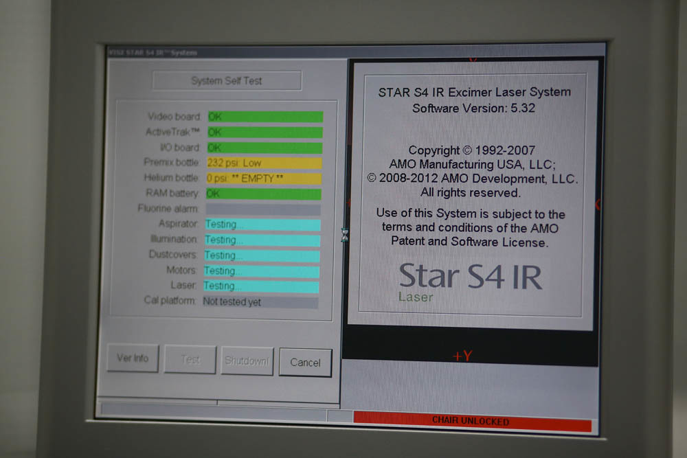
Testing:
The marking for the aiming is visible here:
Activated by pressing the key on the upper left of the joystick. It is positioned on the cornea of the patient during the operation - it is also a “blinking red dot” (its intensity can be changed) during the excimer laser correction operation:

There are three buttons in the left row. The top left includes a red line that is projected onto the patient's face for laying in a strictly longitudinal position without lateral displacement. On the side of the head end there is a label for positioning at eye level. In this way, an accurate styling of the head along the XY axis occurs. The vacuum cushion fixes the head tightly enough in the chosen position. From the left to the right eye, the position is changed by pressing the button with an assistant on the bed of the bed. Next, the surgeon proceeds to centering the eye under a microscope.
The left lower control button for capturing the image of the iris is the control button for the iris recorder (controls the XY position). The left middle one is the cyclothoria control button (by rotating the eyeball around its axis when the patient changes the position from vertical to horizontal).
On the right is the illumination regulation button of the operative field - direct and side illumination. Often, the surgeon almost completely turns off the illumination of the surgical field, since the camera tracking the individual card of the iris works in the infrared range and the “pattern” of the iris and pupil in the dark is determined by the device more quickly.
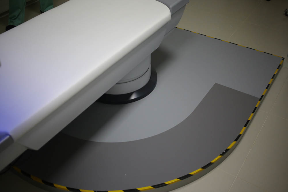
Here is the basic position of the table during the operation:
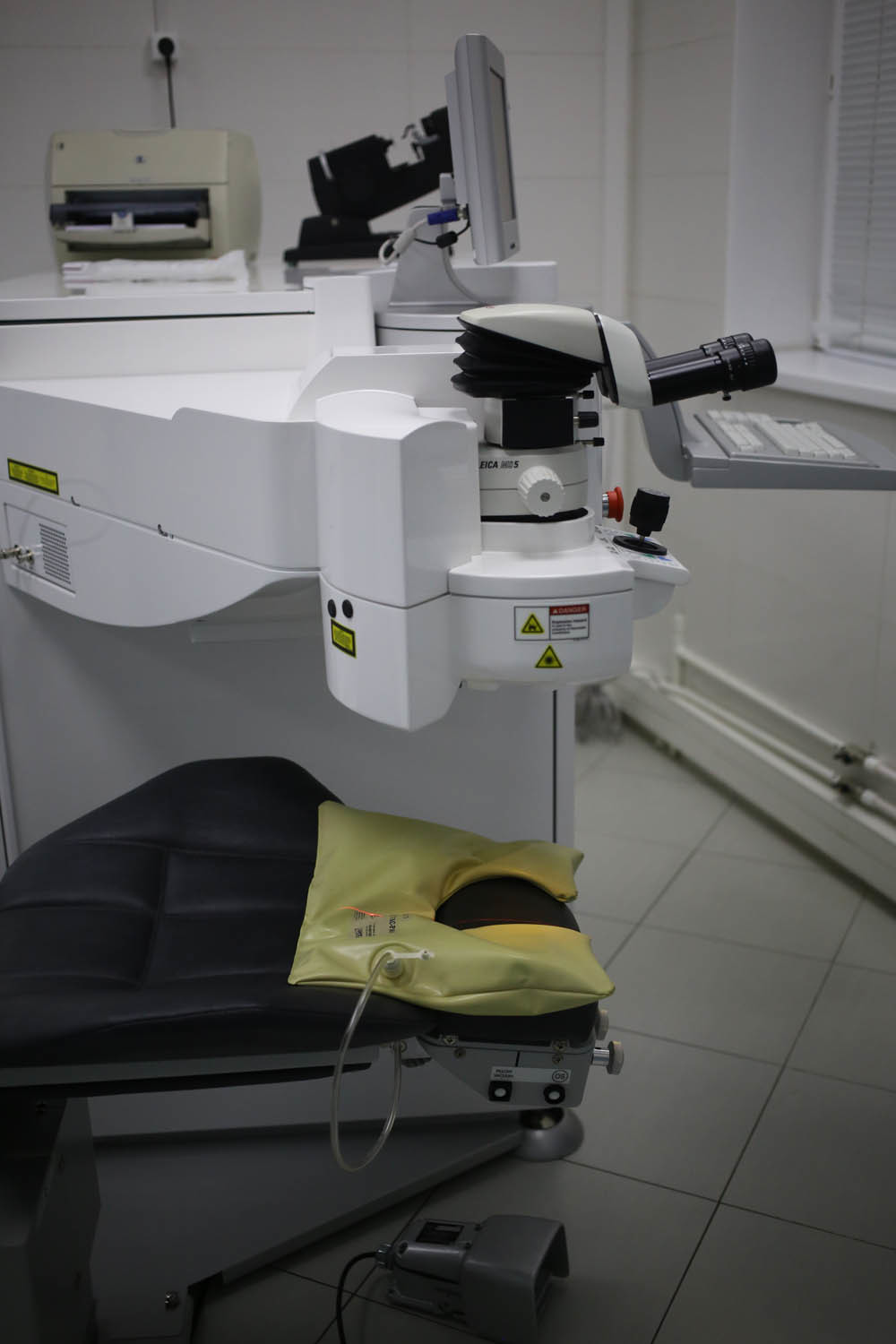
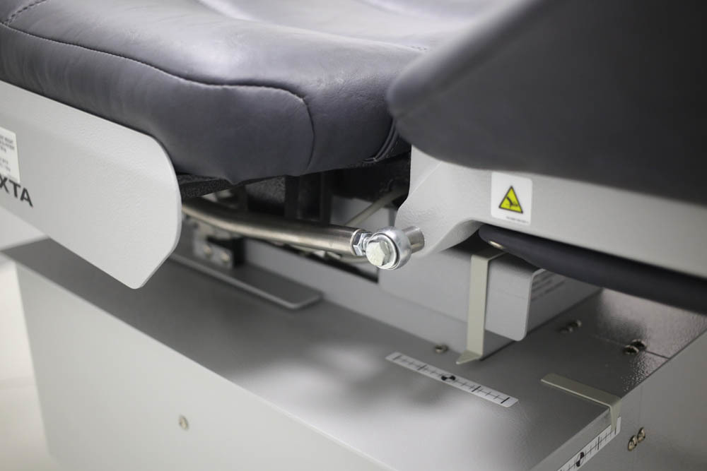
It adjusts in height and other parameters: The
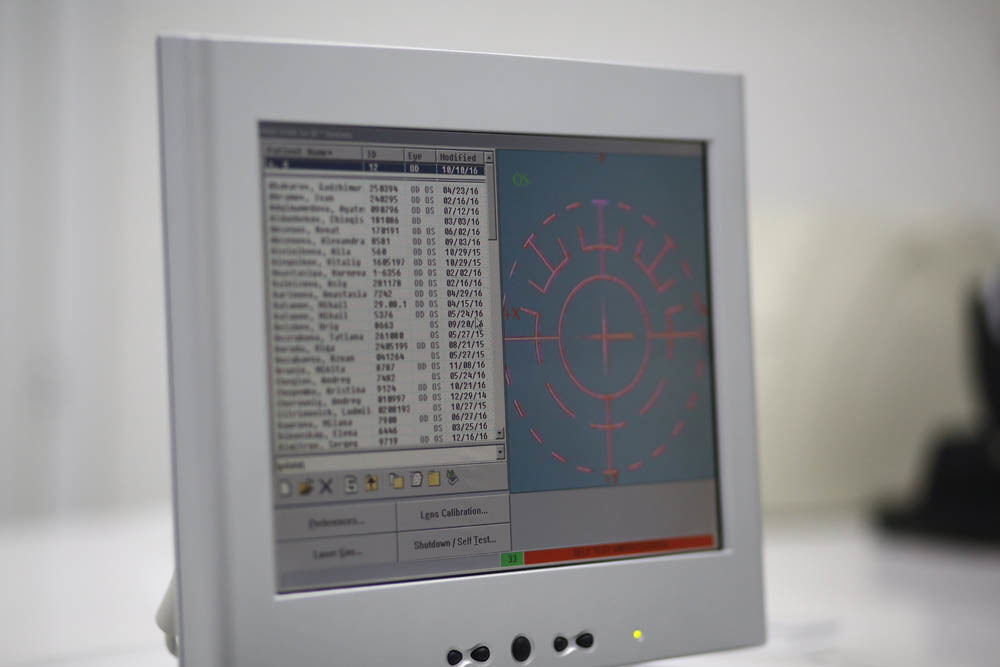
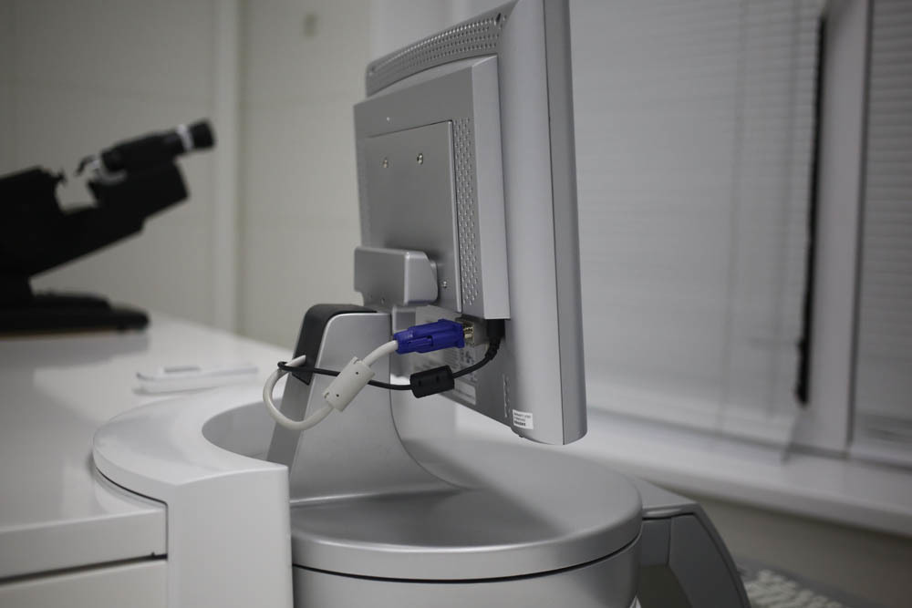
screen on the laser is completely normal:
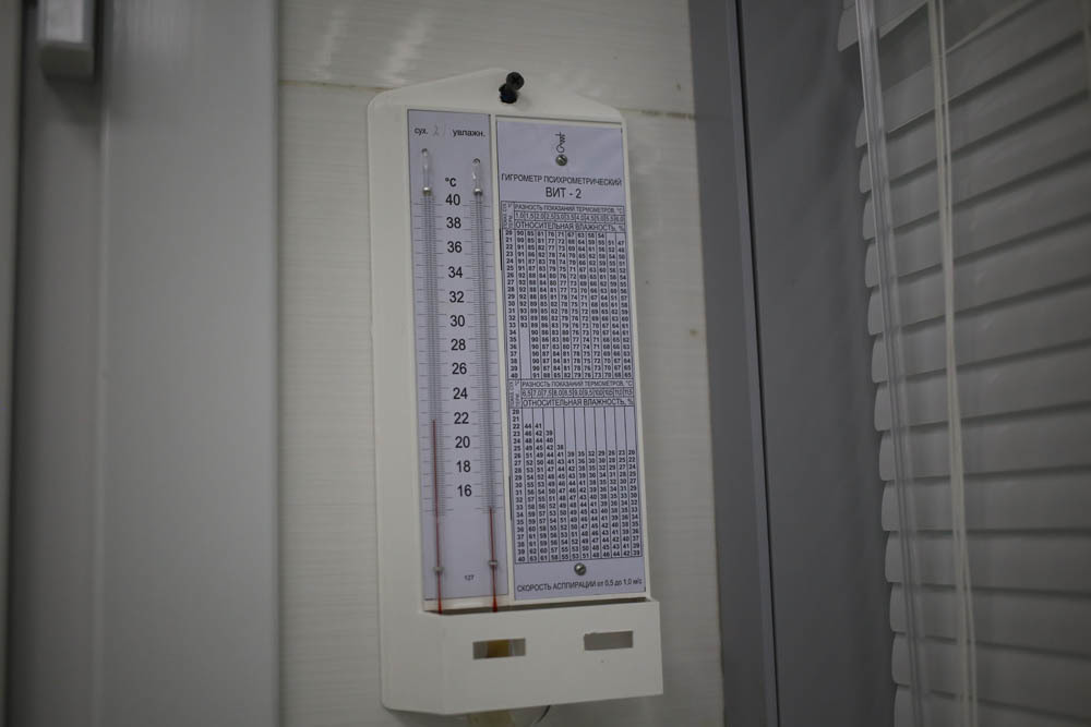
Excimer lasers are extremely sensitive to changes in temperature and humidity in the operating room. Also, no ether and alcohol solutions in the excimer laser operating room should not be. Put constant monitoring of thermometers and hygrometers.
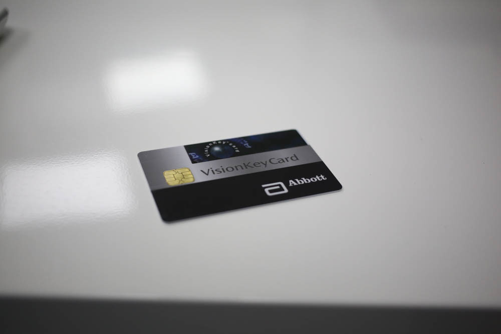
License Card:
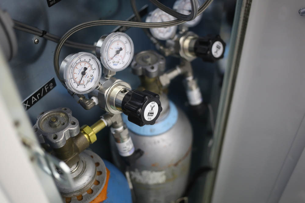
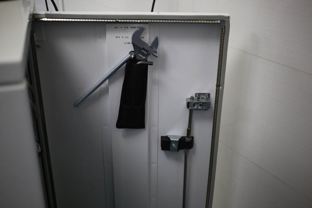
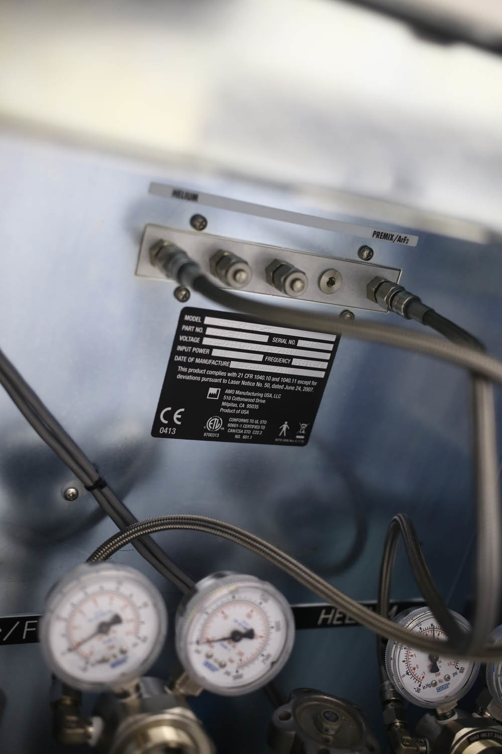
Laser Gas Cylinders:
Assistant screen:
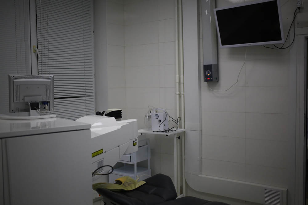
Everything is finished with excimer. Go to the next operating room, where there is a femtolaser VisuMax.It is used to form a flap with femtoLASIK (then the patient goes to the operating room described above under the excimer), for FLEX and SMILE laser correction (then you don’t need to go anywhere - everything is done under one laser), as well as for any corneal incisions, for example, for intracorneal rings with keratoconus). In the case of a general manufacturer of femtolaser and excimer laser systems, in some cases they use one bed for two devices, then they move it in the process of performing different stages.
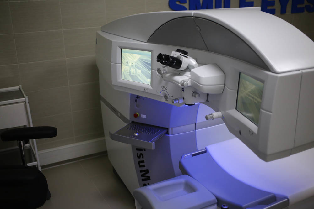
So, this is VisuMax:

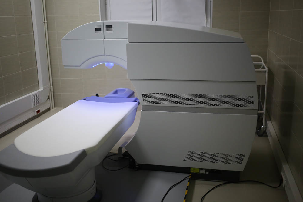
The laser was created to impress with a view - Zeis ordered ergonomics at the design bureau of Mercedes (this laser had both design and cost like Maybach, they not only made the controls very comfortable, but also worked with the appearance). This was motivated very simply: if the patient feels that he is sitting in “Maybach,” then he is somehow more calm than when landing in something from the films about “Alien.”
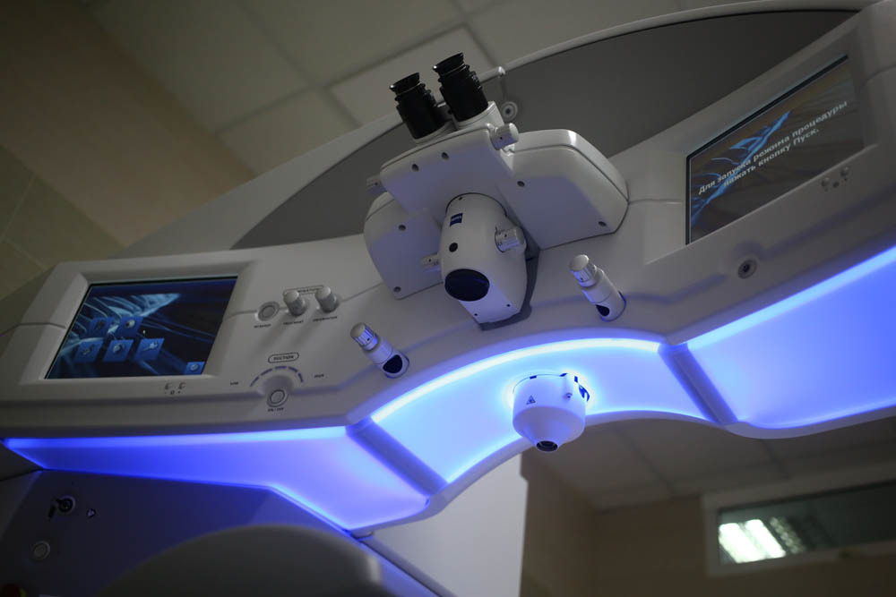
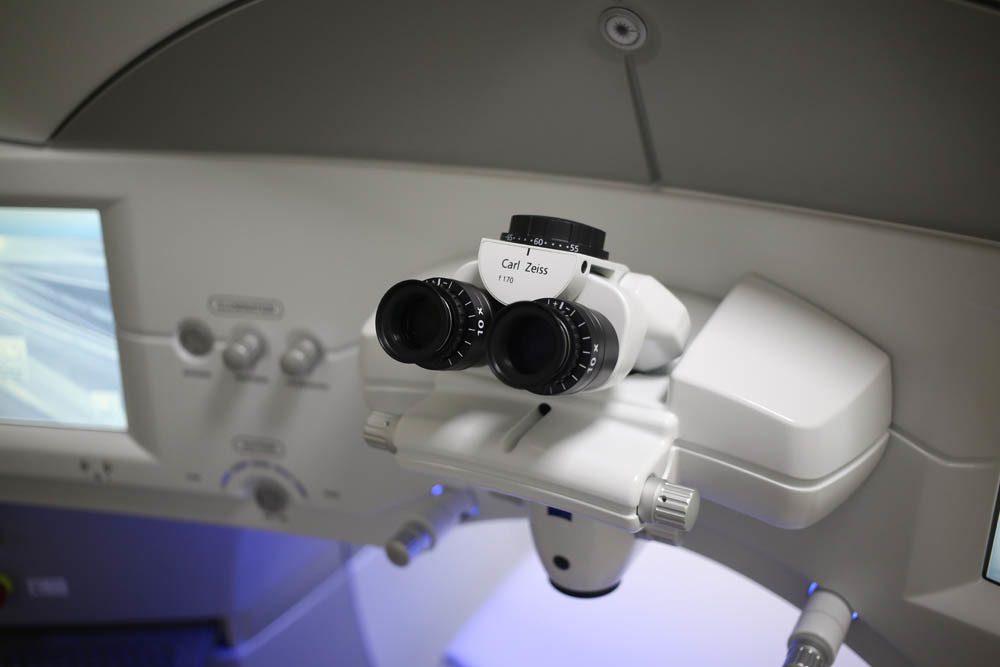
Ergonomics is really great, everything is at hand (or rather, hands): The
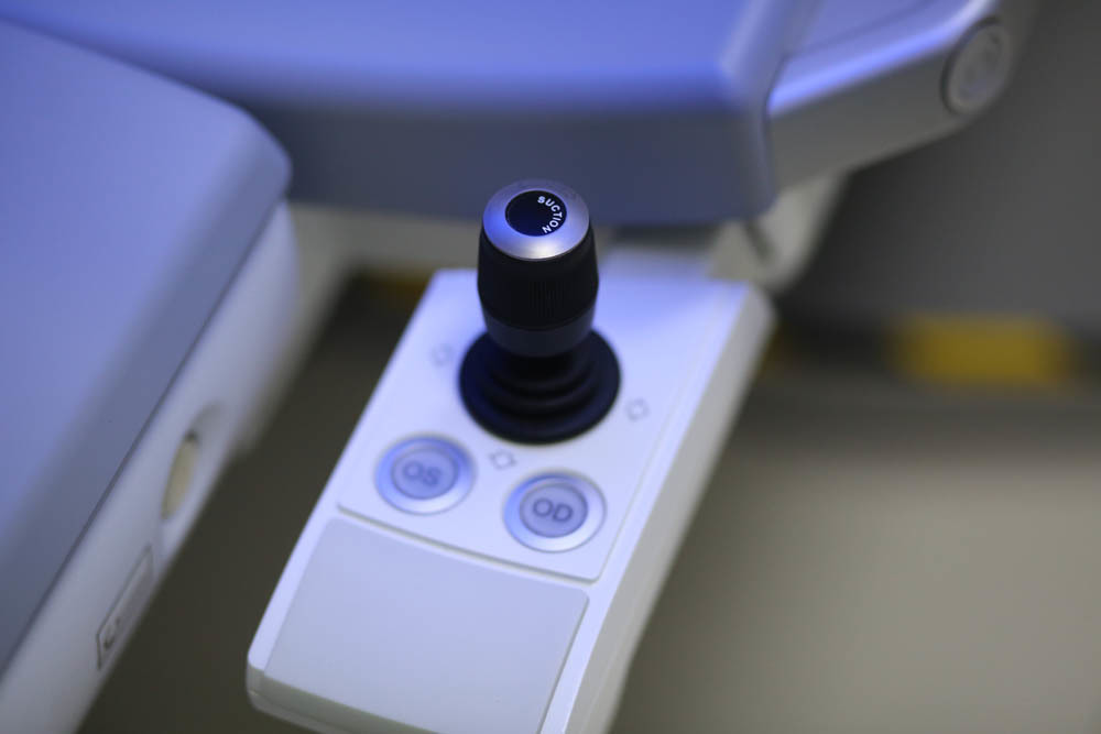
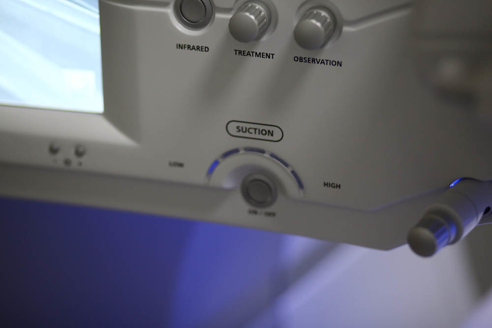
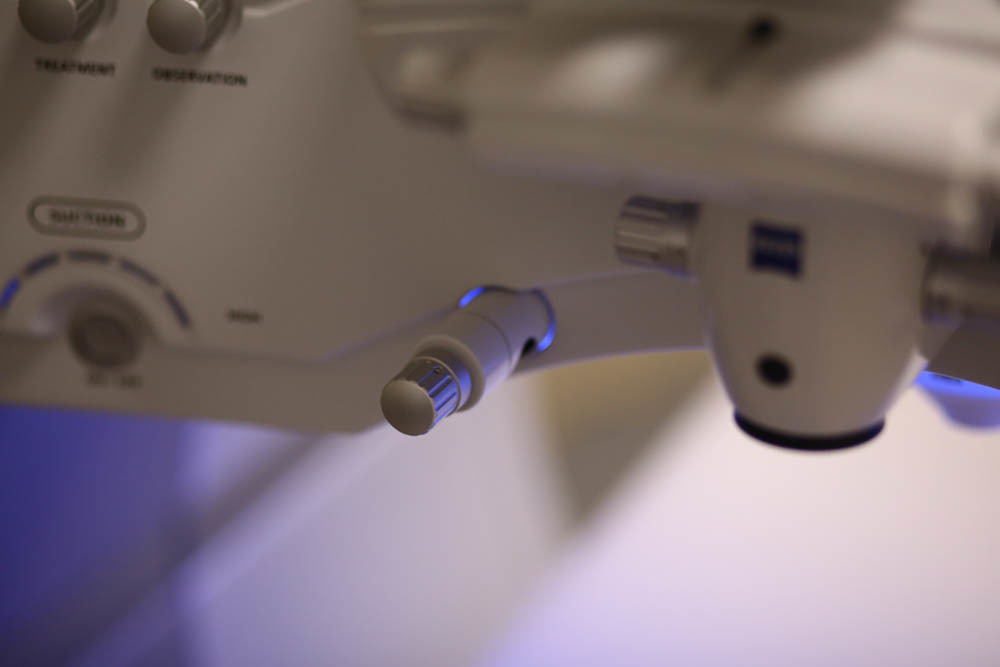
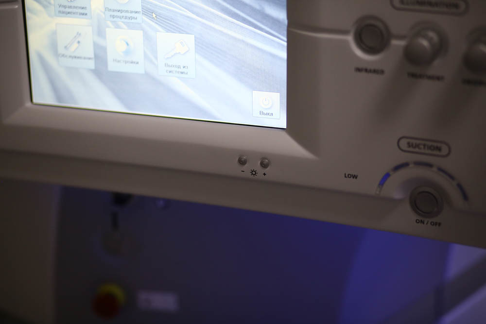
keyboard is almost not needed here, touch screens:
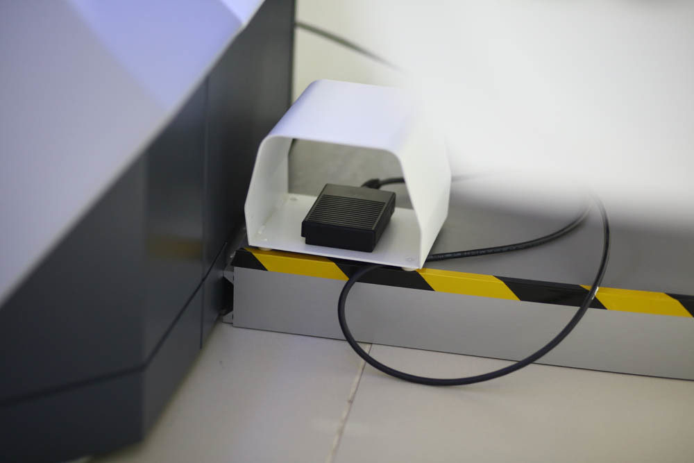
The pedal is slightly different. From the moment it is pressed, a license for a specific operation is considered to be used: The
landing is as follows:
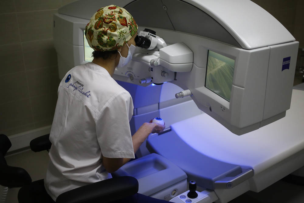
Pay attention to how everything is at hand.
As for the human factor - for a successful surgery there are a couple of important points:
- The surgeon must have stereoscopic, that is, three-dimensional vision, since it is important to see three-dimensional here - the cut is curvilinear, it has depth.
- It is necessary to focus on the sound and light indication - each mode of the laser and each situation have their own indication.
- This is the type of surgery when at the time of surgery the surgeon is left alone with the patient - at this moment only he controls his behavior.
Further - about the software. The interfaces of VisuMax are slightly newer than on excimer, but still on the “ATM” Windows.
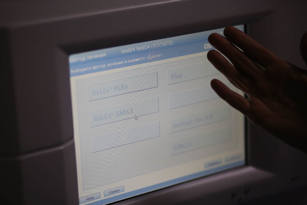
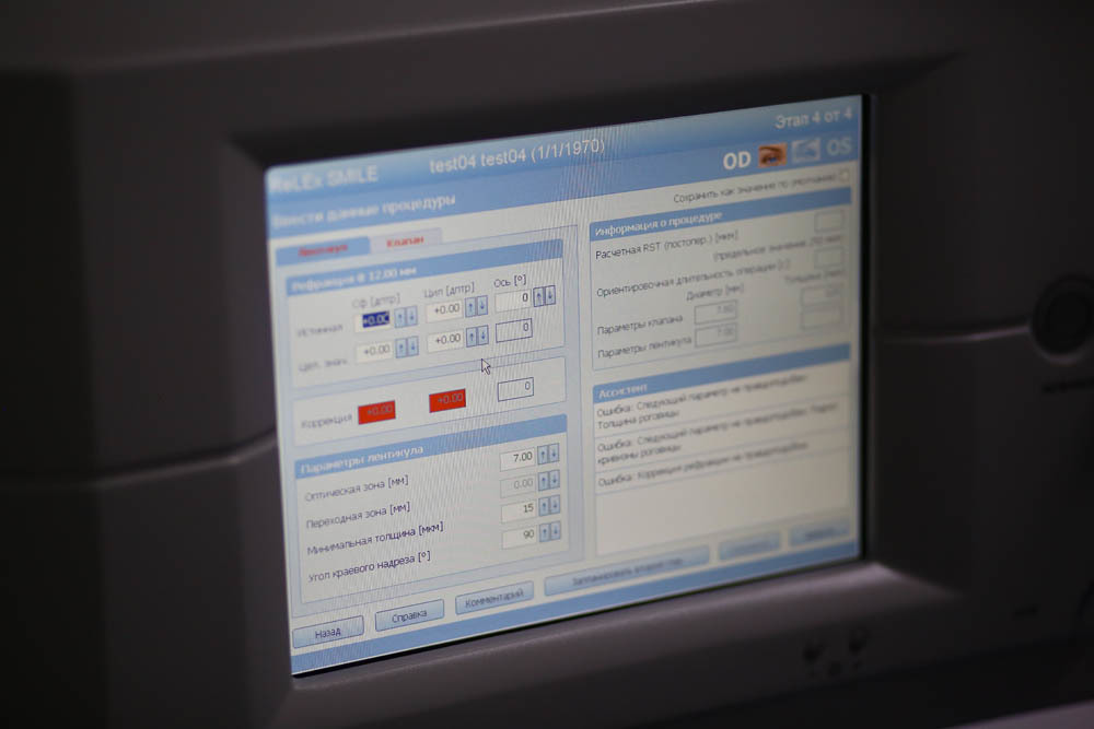
During adjustment, an animation on the left of the screen shows how the cuts will be placed in the cornea and their geometry.
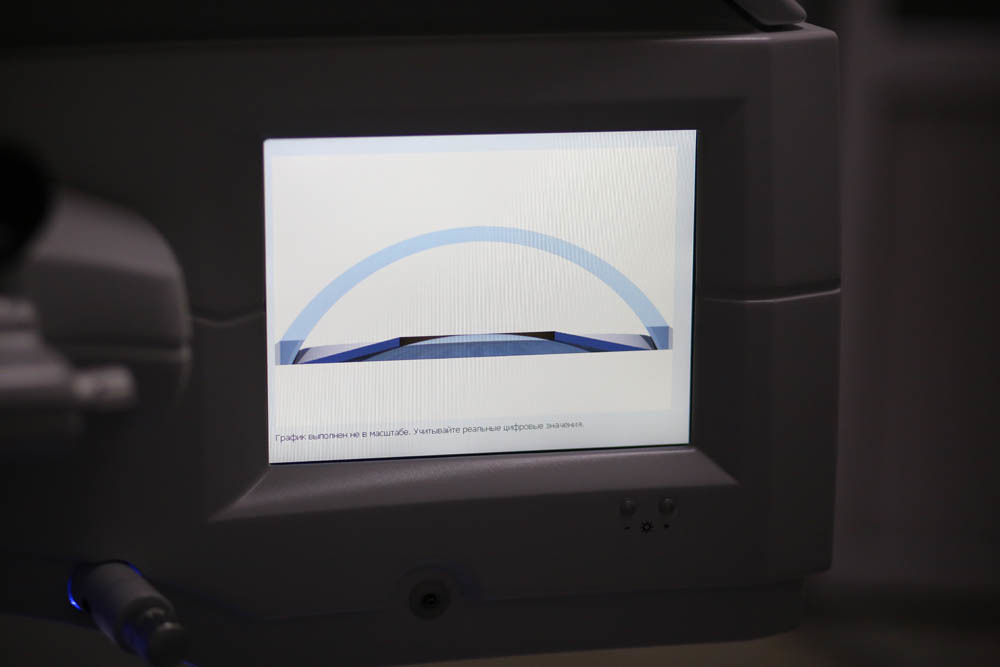
The laser insures against errors: if something in the aggregate of parameters falls out of best practice, warnings are issued. And yes, of course, the last 100 operations are written in the “black box” with all the telemetry and video.
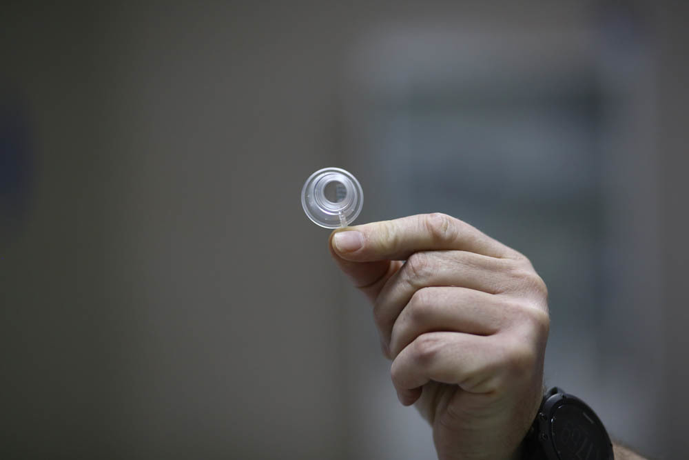
But the cone that comes with the license (this is the replaceable part adjacent to the eye):
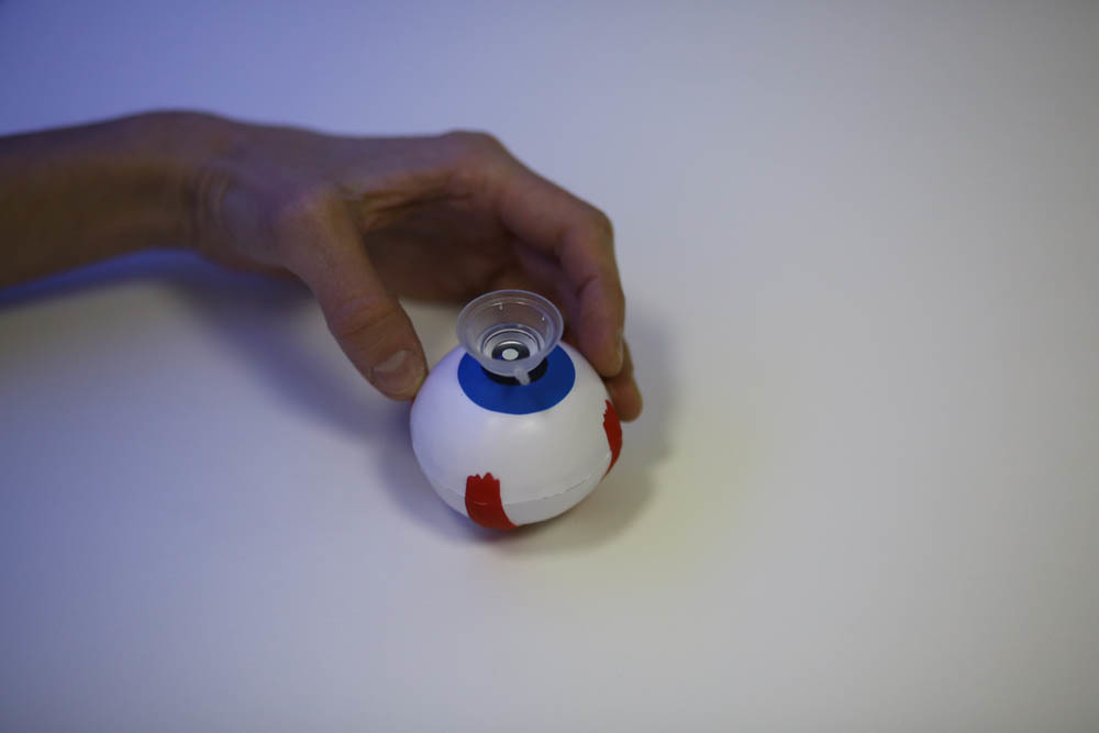
This is how it gets up during the operation (by the way, this is a soft toy that we give to the patient in hand to divert motor skills):
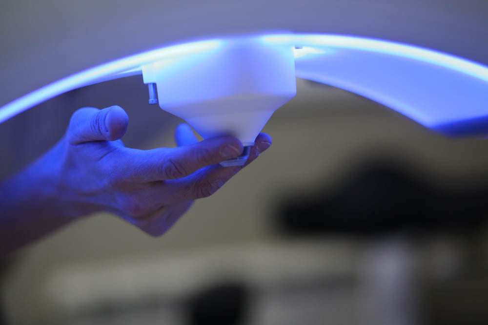
This is the cone located on the laser:
While all. In the first postYou can read about how the Relex SMILE femtosecond laser works at the physical level and how the cut is done inside the cornea, in the second one you can look at the telemetry of a real operation , in the third - what operations are there , in the fourth - different issues about vibration protection, what happens if you shake and so on, in the fifth specifically the FAQ about ReLEx SMILE in Russia, and in the sixth about side effects and diagnostics . This is the seventh, I hope you now have a holistic view of how it all looks. Then we will talk about other methods of correction (in particular, about intraocular lenses), comparison of contact lenses and correction, and a little later I will talk about other eye diseases.