In the Oxford Museum collected portraits of insects from tens of thousands of macro photos
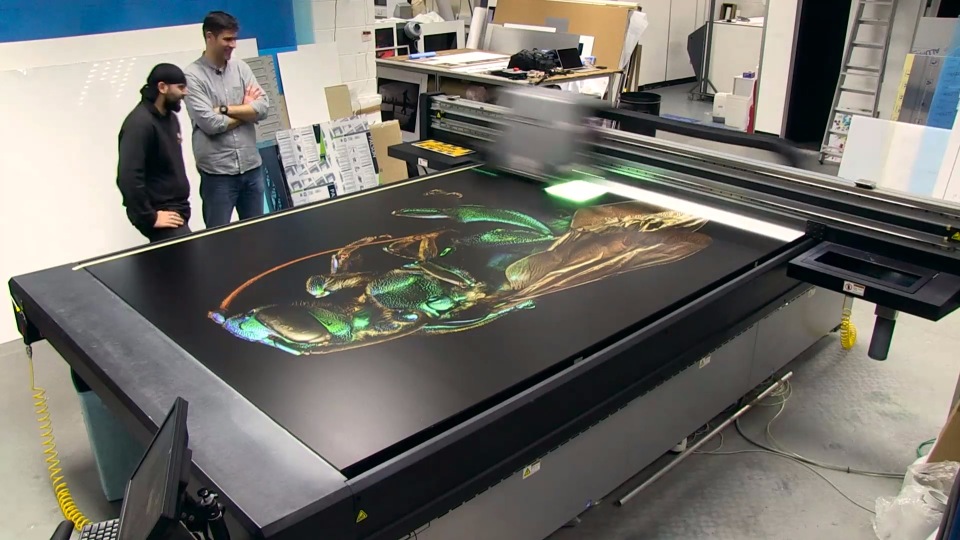
In the spring of this year, the British photographer Levon Biss, together with the Oxford Museum of Natural History, presented the “Microsculpture” project . Looking at the museum, its visitors are surrounded by three-meter portraits of microscopic insects. In addition to the incredible beauty, these photos are of scientific value. Often, many insects do not exceed one centimeter in length, and all the subtle details, the "microsculpture" of the body are hidden from the human eye. It is not surprising that this particular entomological term served as the name for a joint project of a scientist and a photographer.
In everyday life, we are accustomed to not paying attention to insects, but many of us in childhood looked at their pictures in encyclopedias: the female praying mantis prepares for breakfast, and here the rhinoceros beetle, breaking all the laws of aerodynamics, rushes over the camera lens. Perhaps, after reading someone, someone wanted to link their life with a photo, but someone had the idea to look at bugs and spiders under a microscope: they became entomologists and devoted their lives to studying these amazing creatures. Usually their life paths rarely overlap, but this is what happens if a talented photographer meets an enthusiastic entomologist.
Until 2014, Levon Byss didn’t think that he would ever take pictures of insects: for almost twenty years he was engaged in shooting portraits for sports and business publications. His six-year-old son, Sebastian, helped Biss to switch to insect microsculptures. The boy spent a lot of time in the garden in search of various bugs, and Levon began shooting them on camera so that his son could see his finds in the smallest detail. The usual macro lens for such filming was not enough, and Byss went on: he came up with another, more labor-intensive method, which gave amazing results.
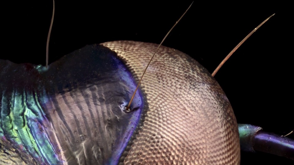
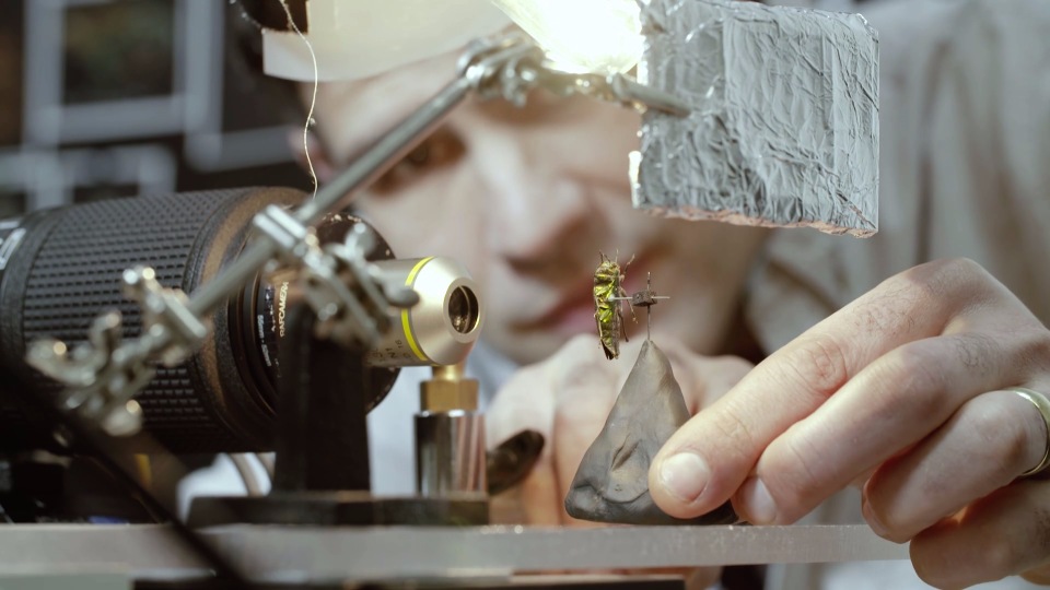
Levon took his 36-megapixel Nikon D810 with a macro lens and attached a microscope eyepiece to it with a tenfold magnification. With this setup, Biss was able to capture the smallest details, but due to the shallow depth of field, each part of the insect was forced to shoot separately. After each flash, the automatically moving tripod slowly lifted the camera, only 10 microns, a distance equal to the length of two red blood cells (red blood cells). For comparison: the average width of a human hair is 75 microns. Thus, 1/20 or 1/30 of the total picture was obtained from several hundred shots. At the same time, it was impossible to allow parts “stitched” together to differ in color and perspective, so the photographer carefully followed the lighting in the frame. To remove one "portrait", it took Levon about 30 hours. Then there were long hours of processing, which took shape in the weeks: Levon collected one image from 8-10 thousand fragments. In total, it took up to three weeks to take, process and retouch photos, but it was worth it: “Until you see them very close - I mean really close,” says Biss, “you don’t think that each of the fine hairs on the wings of an ordinary fly has unique outlines, or on the back of a ground beetle dimples of an ideal geometric shape. "
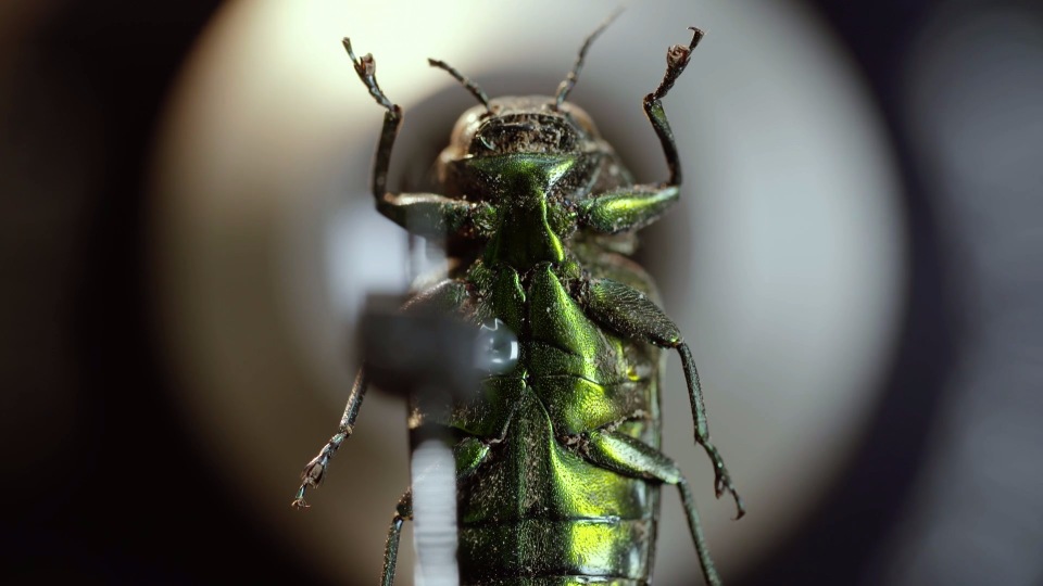
After the first successful experiments, Levon Byss went to the Oxford Museum of Natural History, where he demonstrated his work to an entomologist, Dr. James Hogan. The latter was so impressed that he offered the photographer access to his collection of five million insects, and personally selected the most interesting of them for shooting. Every few months, the entomologist prepared more than a dozen impressive specimens, many of which had unique features of the structure. The horse beetles , which have become the objects of the exhibition, have hexagonal dimples all over their backs that disperse the water. This seemingly uncomplicated structure helps the racers living on land to survive the floods. At the same time, comically tiny wings and long legs of a flightless mothfrom the island of Marion allow it to quietly exist in the cold climate of the southern Indian Ocean.
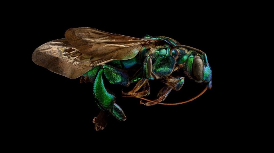
In the process of working on a project, Levon Biss discovered many interesting patterns for himself. He was especially surprised when he learned that the color of an insect seems so bright not because of the pigment that could fade over time, but because of the refraction of light from their delicately textured bodies. “I worked on a woodworm beetle of extraordinary coloring, which was caught by A.R. Wallace - a contemporary of Darwin. This beetle is already 160 years old, and it is still full of color, ”the photographer notes.
The Union of Art and Science opened the door to an exciting, full of beauty world of unusual insects. The tangled shapes, colors, microsculpture of bodies, which used to be available only at the microscopic level, are now open to the eyes of millions of people. The photos are exhibited in the museum until October 30, but you can also study or purchase any of the exhibits in detail without leaving your home: the fruits of Levon Biss and James Hogan’s teamwork are available free of charge on the project’s official website. There you can see each of the 22 copies, like under a microscope - for visitors there is the possibility of scaling the image.