3D printing of protein molecule models
So, the fashion for 3D printing has reached our modest institution, proudly called the Laboratory of Molecular Biology . A couple of weeks ago, the workshops bought a 3D printer, which initiated a heated discussion among biologists - how a new thing could be useful. Physiologists, for example, have already gathered to print some complicated details for a new mechanism designed to test all sorts of behavioral reactions in rats. We are engaged in determining the three-dimensional structure of the protein by x-ray diffraction analysis (and, for some time, cryo-electron microscopy) and for us the benefits of 3D printing are not so obvious. Accordingly, we rummaged through the Internet to understand what people are printing in terms of protein structures. The resulting reviewer with beautiful pictures and video of printed models is under the cut.
As Comrade Friedrich Engels rightly pointed out more than a hundred years ago, "life is a way of existence of protein bodies." That is, our bodies are largely made of protein and proteins perform the most important functions in our body - structural (the shape of the cells rests on the protein cytoskeleton), motor (muscles contract due to the cunning work of motor proteins), enzymatic (say, food is digested due to protein-enzymes ), transport (oxygen from the lungs to the tissues carries the hemoglobin protein) and so on; in short, without protein - nowhere.
Accordingly, it is very interesting to know how proteins work. From the point of view of a biochemist, protein is such a black box. You can give him different substances at the entrance and then watch what happens at the exit (at what speed, etc.). The definition of the three-dimensional structure of a protein molecule in a sense opens this black box, the structure of the protein is a diagram of how the black box is arranged inside. It is not always possible from the structure to immediately understand how the protein works, but the structure, of course, opens our eyes and allows us to plan further experiments.
A protein is a very large molecule (macromolecule). From a chemical point of view, it is a linear polymer, from 70 to more than 1000 amino acid monomers in length. Due to the different lateral groups of amino acids, a long protein molecule collapses into a compact globule, each protein has its own styling. It is the structure of folded protein that scientists determine. The leading method is x-ray diffraction analysis, which allows you to calculate the coordinates of the atoms of the protein by x-ray diffraction on the crystal of the corresponding protein. Crystallographers have a convention - any specific protein structure must be uploaded to the Protein Data Bank), in which the number of structures has already exceeded 100,000. Accordingly, all structures are in the public domain, they can be downloaded, viewed, printed on a 3D printer, and so on. I wrote in more detail about protein structures on Habré earlier; in a more detailed form about structures can be read here .
It should be noted that the task of displaying the protein structure is not so simple. A protein consists of several thousand atoms, which, at first glance, seem like a terrible mess (“a” and “c” in the picture). For different tasks, different mappings are used. For example, if we want to understand how the main chain of a protein is laid, then we want to display the protein in a schematic form, where only the main chain path (“g” in the picture) will be indicated using special notation — spiral to denote alpha-helices, arrows for designations of beta-folded sheets ("e", "e" in the picture).
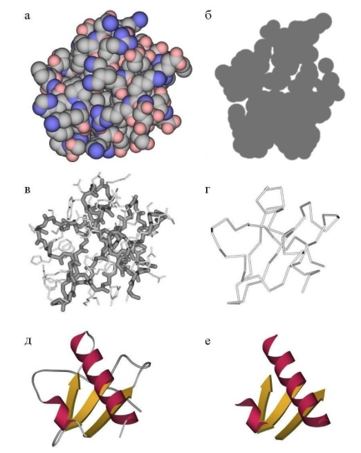
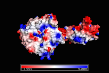
If we want to see the protein as a voluminous body, we can display the surface of the protein (then it becomes like a potato); the surface can be painted using different parameters. The most useful is to colorize according to the electrostatic potential, then the charged areas will be immediately visible.
If we want to understand how a drug molecule or chemical substrate binds to our protein, then we display only a small part of the protein — the active center of the enzyme, a small number of key amino acids that interact directly with the drug.
For starters, some entertaining videos.
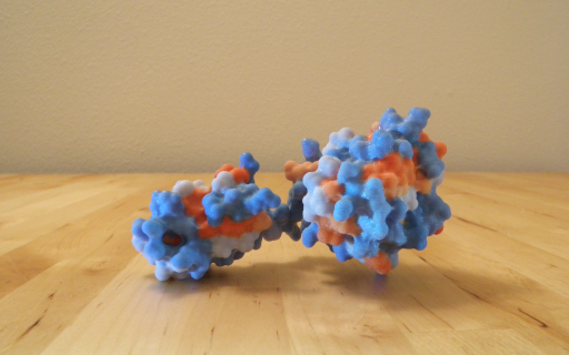
Here is a very vivid demonstration of how many molecules of actin protein are assembled into the so-called “actin filaments”, which form the cytoskeleton, on which the whole form of our cells rests, plus it also works in the muscles. Individual actin molecules are printed on a 3D printer, the surface is painted according to electrostatic potential.
Here's something cooler: a hemoglobin molecule that carries oxygen in our body (red blood cells are packed with this protein). Hemoglobin consists of four subunits (individual protein molecules), plus a small heme molecule bound to hemoglobin, which carries an iron atom, which binds oxygen. In the manufacture of this model used more complex tech. the process and, as a result, the transparent external surface reflects the real surface of the protein molecule, and inside, under the transparent external layer, multi-colored alpha helices and other elements of the secondary structure are visible, clearly showing the course of the main chain of the protein. The video shows the assembly of a whole hemoglobin from four subunits, the subunits are mounted on magnets. At the very end, a small heme molecule is pushed into place.
Here is another video of the same model, here it shows the secondary structure better and shows how lowering the model into water allows you to see the secondary structure in more contrast. Another difference - in this model, the outer layer of the protein is not just transparent, as in the previous version, but also slightly tinted by the electrostatic potential:
These videos were taken from the channel of the Japanese 3D printing enthusiast Kawakami Masaru, more videos with different printed protein structures can be found here:
http://www.youtube.com/channel/UCsrgChR36VUMVy8GejyuD0Q/videos
Of course, smart businessmen couldn't help but pick up the idea and the production of molecular beauty has already been put on stream: such cognitive structures of protein can already be bought .

Again, if you want to print your protein, it is not necessary to buy a 3D printer. There are already a sufficient number of companies selling both ready-made kits (for example, three-dimensional DNA models) and printing any kind of protein to order. For example, 3D Molecular Designs .
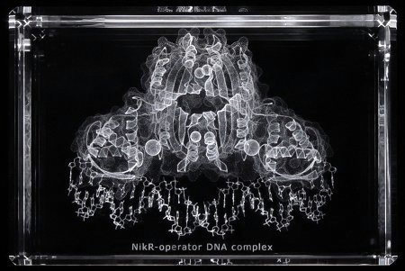
On the question of business: in addition to 3D printing itself, there have long been companies ready to print the three-dimensional structure of your favorite protein in a transparent crystal . The idea is quite in demand among structural biologists: for example, a similar crystal was presented by a chef to one of my acquaintances after she determined the first protein structure in her life. The scientific value of such a crystal tends to zero, but it looks very beautiful and aesthetically pleasing.
If someone thinks that this is all jokes and mischief - not at all. The seriousness of the situation is shown by the recently launched repository of ready-made 3D models from the American NIH- National Institutes of Health in collaboration with the National Library of Medicine (these are the most serious structures that are part of the US Department of Health). In addition to protein structures of high (X-ray crystallography) and low resolution (cryo-electron microscopy), the repository also shows models of tube stands and other laboratory details. The repository was created, as I understand it, to improve the internal exchange of models in NIH. Now it has 452 models.
If you were inspired by the above pictures and there is access to a 3D printer, then printing the protein structure itself is quite easy. There are many step-by-step instructions on the Internet, here are two of the most sensible and detailed:
http://www.instructables.com/id/3D-Print-a-Protein-Modeling-a-Molecular-Machine/?ALLSTEPS
http: // www. over-engineered.com/projects/3d-printed-protein
In short, you must first install one of the programs for displaying protein structures . Then - find the structure to print. All structures are located in Protein Data Bank , there is a search for keywords. In addition to the search, they have a marvelous project “Molecule of the Month”- they choose a certain protein and talk in detail about its structure and function for a wide audience of non-professionals. The main page of the project looks awesome because of the abundance of obscure names of proteins, but if you start clicking on the links, there will be beautiful pictures and clear descriptions of the work and structure of a protein. I hope this information helps you select the protein to print.
After loading the structure (the Protein Data Bank has a wonderful “Download files” button) and displaying it in the appropriate program, you need to choose in which form you want to print the molecule. In the above instructions, it is proposed to display the protein as a surface painted according to electrostatic potential, export the surface, convert it to a print format and - voila! A potato-like volumetric protein model is really easy to print.
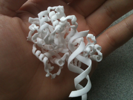
However, it would be interesting to print and, say, a schematic representation of the main chain of the protein, something like this (here, as far as I see, the complex of protein and DNA):
The only thing is that such a skeletal structure of the protein can be fragile. For such printing, it is recommended to use a program to display Chimera protein structures.because it can export directly .stl files that are understood by most 3D printers. Accordingly, any type of molecule of your choice in Chimera is quietly exported and can be printed, including skeletal representation. Here is a short video presentation explaining this process a bit:
As we have just seen, people are actively printing protein structures. The question remains open: why are they doing this? So far, the only answer is for educational, demonstration, educational purposes. Proteins are complex three-dimensional objects and the ability to hold such a "molecule" in the hand, of course, helps to understand how the protein is built and works. Plus, many proteins are simply visually beautiful. Printing the skeletal structure of a protein can be quite a serious technical challenge, and then an impressive demonstration of the capabilities of 3D printing. So if you have a 3D printer, free time and enthusiasm ... you know what to do!
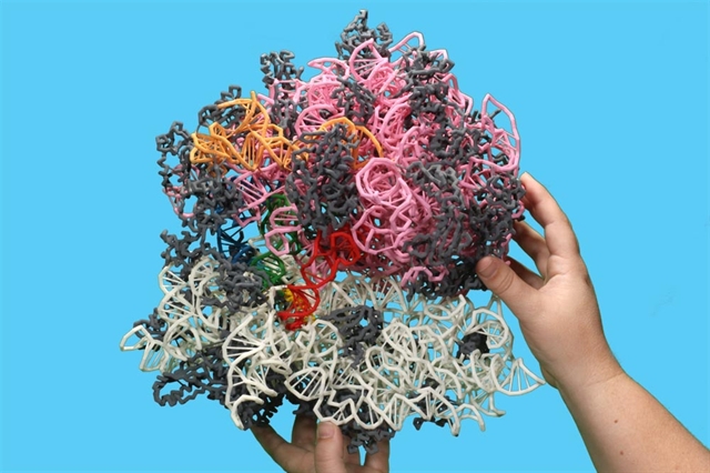
This beauty is a ribosome .
Briefly about protein structures
As Comrade Friedrich Engels rightly pointed out more than a hundred years ago, "life is a way of existence of protein bodies." That is, our bodies are largely made of protein and proteins perform the most important functions in our body - structural (the shape of the cells rests on the protein cytoskeleton), motor (muscles contract due to the cunning work of motor proteins), enzymatic (say, food is digested due to protein-enzymes ), transport (oxygen from the lungs to the tissues carries the hemoglobin protein) and so on; in short, without protein - nowhere.
Accordingly, it is very interesting to know how proteins work. From the point of view of a biochemist, protein is such a black box. You can give him different substances at the entrance and then watch what happens at the exit (at what speed, etc.). The definition of the three-dimensional structure of a protein molecule in a sense opens this black box, the structure of the protein is a diagram of how the black box is arranged inside. It is not always possible from the structure to immediately understand how the protein works, but the structure, of course, opens our eyes and allows us to plan further experiments.
A protein is a very large molecule (macromolecule). From a chemical point of view, it is a linear polymer, from 70 to more than 1000 amino acid monomers in length. Due to the different lateral groups of amino acids, a long protein molecule collapses into a compact globule, each protein has its own styling. It is the structure of folded protein that scientists determine. The leading method is x-ray diffraction analysis, which allows you to calculate the coordinates of the atoms of the protein by x-ray diffraction on the crystal of the corresponding protein. Crystallographers have a convention - any specific protein structure must be uploaded to the Protein Data Bank), in which the number of structures has already exceeded 100,000. Accordingly, all structures are in the public domain, they can be downloaded, viewed, printed on a 3D printer, and so on. I wrote in more detail about protein structures on Habré earlier; in a more detailed form about structures can be read here .
It should be noted that the task of displaying the protein structure is not so simple. A protein consists of several thousand atoms, which, at first glance, seem like a terrible mess (“a” and “c” in the picture). For different tasks, different mappings are used. For example, if we want to understand how the main chain of a protein is laid, then we want to display the protein in a schematic form, where only the main chain path (“g” in the picture) will be indicated using special notation — spiral to denote alpha-helices, arrows for designations of beta-folded sheets ("e", "e" in the picture).
If we want to see the protein as a voluminous body, we can display the surface of the protein (then it becomes like a potato); the surface can be painted using different parameters. The most useful is to colorize according to the electrostatic potential, then the charged areas will be immediately visible.
If we want to understand how a drug molecule or chemical substrate binds to our protein, then we display only a small part of the protein — the active center of the enzyme, a small number of key amino acids that interact directly with the drug.
Printed Protein Structures
For starters, some entertaining videos.
Here is a very vivid demonstration of how many molecules of actin protein are assembled into the so-called “actin filaments”, which form the cytoskeleton, on which the whole form of our cells rests, plus it also works in the muscles. Individual actin molecules are printed on a 3D printer, the surface is painted according to electrostatic potential.
Here's something cooler: a hemoglobin molecule that carries oxygen in our body (red blood cells are packed with this protein). Hemoglobin consists of four subunits (individual protein molecules), plus a small heme molecule bound to hemoglobin, which carries an iron atom, which binds oxygen. In the manufacture of this model used more complex tech. the process and, as a result, the transparent external surface reflects the real surface of the protein molecule, and inside, under the transparent external layer, multi-colored alpha helices and other elements of the secondary structure are visible, clearly showing the course of the main chain of the protein. The video shows the assembly of a whole hemoglobin from four subunits, the subunits are mounted on magnets. At the very end, a small heme molecule is pushed into place.
Here is another video of the same model, here it shows the secondary structure better and shows how lowering the model into water allows you to see the secondary structure in more contrast. Another difference - in this model, the outer layer of the protein is not just transparent, as in the previous version, but also slightly tinted by the electrostatic potential:
These videos were taken from the channel of the Japanese 3D printing enthusiast Kawakami Masaru, more videos with different printed protein structures can be found here:
http://www.youtube.com/channel/UCsrgChR36VUMVy8GejyuD0Q/videos
Of course, smart businessmen couldn't help but pick up the idea and the production of molecular beauty has already been put on stream: such cognitive structures of protein can already be bought .
Again, if you want to print your protein, it is not necessary to buy a 3D printer. There are already a sufficient number of companies selling both ready-made kits (for example, three-dimensional DNA models) and printing any kind of protein to order. For example, 3D Molecular Designs .
On the question of business: in addition to 3D printing itself, there have long been companies ready to print the three-dimensional structure of your favorite protein in a transparent crystal . The idea is quite in demand among structural biologists: for example, a similar crystal was presented by a chef to one of my acquaintances after she determined the first protein structure in her life. The scientific value of such a crystal tends to zero, but it looks very beautiful and aesthetically pleasing.
If someone thinks that this is all jokes and mischief - not at all. The seriousness of the situation is shown by the recently launched repository of ready-made 3D models from the American NIH- National Institutes of Health in collaboration with the National Library of Medicine (these are the most serious structures that are part of the US Department of Health). In addition to protein structures of high (X-ray crystallography) and low resolution (cryo-electron microscopy), the repository also shows models of tube stands and other laboratory details. The repository was created, as I understand it, to improve the internal exchange of models in NIH. Now it has 452 models.
How to print the protein structure yourself
If you were inspired by the above pictures and there is access to a 3D printer, then printing the protein structure itself is quite easy. There are many step-by-step instructions on the Internet, here are two of the most sensible and detailed:
http://www.instructables.com/id/3D-Print-a-Protein-Modeling-a-Molecular-Machine/?ALLSTEPS
http: // www. over-engineered.com/projects/3d-printed-protein
In short, you must first install one of the programs for displaying protein structures . Then - find the structure to print. All structures are located in Protein Data Bank , there is a search for keywords. In addition to the search, they have a marvelous project “Molecule of the Month”- they choose a certain protein and talk in detail about its structure and function for a wide audience of non-professionals. The main page of the project looks awesome because of the abundance of obscure names of proteins, but if you start clicking on the links, there will be beautiful pictures and clear descriptions of the work and structure of a protein. I hope this information helps you select the protein to print.
After loading the structure (the Protein Data Bank has a wonderful “Download files” button) and displaying it in the appropriate program, you need to choose in which form you want to print the molecule. In the above instructions, it is proposed to display the protein as a surface painted according to electrostatic potential, export the surface, convert it to a print format and - voila! A potato-like volumetric protein model is really easy to print.
However, it would be interesting to print and, say, a schematic representation of the main chain of the protein, something like this (here, as far as I see, the complex of protein and DNA):
The only thing is that such a skeletal structure of the protein can be fragile. For such printing, it is recommended to use a program to display Chimera protein structures.because it can export directly .stl files that are understood by most 3D printers. Accordingly, any type of molecule of your choice in Chimera is quietly exported and can be printed, including skeletal representation. Here is a short video presentation explaining this process a bit:
Conclusion (or what for goat bayan)
As we have just seen, people are actively printing protein structures. The question remains open: why are they doing this? So far, the only answer is for educational, demonstration, educational purposes. Proteins are complex three-dimensional objects and the ability to hold such a "molecule" in the hand, of course, helps to understand how the protein is built and works. Plus, many proteins are simply visually beautiful. Printing the skeletal structure of a protein can be quite a serious technical challenge, and then an impressive demonstration of the capabilities of 3D printing. So if you have a 3D printer, free time and enthusiasm ... you know what to do!
This beauty is a ribosome .