Overview of the tasks of computer vision in medicine

Computer vision and machine learning find their application in many areas of human activity. Medicine was no exception. This article discusses the most interesting, in the opinion of the author, tasks of computer vision in medicine.
Automatic detection of circulating tumor cells
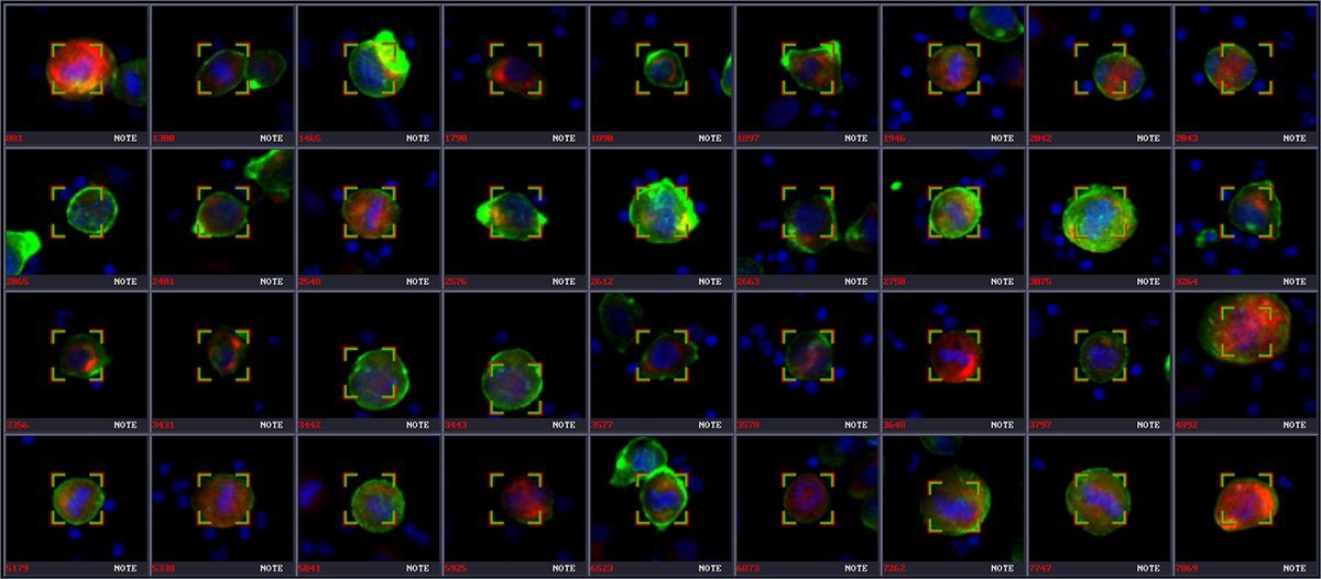
Circulating tumor cells interbreeding with several fluorescent antibodies.
Circulating tumor cells are cells that separate from the site of the main tumor and spread through the bloodstream, forming secondary tumors in other organs.
Early detection of such cells and assessment of disease progression is very important for effective treatment, therefore, systems for automatic detection of tumor cells are being actively developed. For example, researchers from Germany [2] obtained accuracy> 99%, recall = 88% and precision = 86% on a small experimental data set.
Sources:
- Automated Detection of Circulating Cells Using Low Level Features
- Automated Detection of Circulating Tumor Cells with Naive Bayesian Classifiers
- bioview.com/applications/circulating-tumor-cells
Automatic detection of diabetic retinopathy

The classification of images depending on the degree of the disease: a) normal; b) mild; c) moderate; d) severe; e) prolific
The authors of the kaggle competition for the detection of diabetic retinopathy claim that 40-45% of Americans with diabetes are also affected by diabetic retinopathy (I think that in Russia the figures are about the same). The progression of visual impairment can be slowed or prevented if the disease is detected in time. Thus, the development of systems for the detection of diabetic retinopathy is also relevant.
The best quadratic weighted kappa result in competitions was 0.84958 (Private Leaderboard). Some researchersDevelop adapters for smartphones for retina shots. They do this with 28D or 40D lenses (such lenses cost about $ 300) and nozzles for a smartphone, which are printed on a 3D printer.
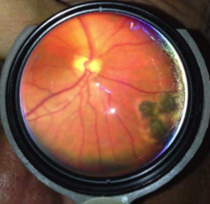
An adapter for retinal images
As you can see, the ability to check the status of your retina without going to the hospital is just around the corner.
Sources:
- www.kaggle.com/c/diabetic-retinopathy-detection
- Algorithms for the Automated Detection of Diabetic Retinopathy Using Digital Fundus Images: A Review
- 3D Printed Smartphone Indirect Lens Adapter for Rapid, High Quality Retinal Imaging
- Smartphone fundoscopy. Ophthalmology
Segmentation of MRI images
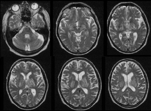
MRI images of the brain
The method of magnetic resonance imaging (MRI) is widely used to diagnose and track the dynamics of brain diseases, as well as to study its functioning. The method allows to obtain three-dimensional images of high quality and resolution, which are built on a set of consecutive two-dimensional "slices". The marking of brain MRI images into anatomical structures is an important step for further analysis in many tasks in this area.
Full marking of a three-dimensional image involves the division (segmentation) of the brain volume into several dozen regions corresponding to the basic anatomical structures. Each point (voxel) is associated with an anatomical structure label. Thus, manual marking in this case becomes a long and laborious process. Therefore, algorithms are needed that automate the anatomical markup process. [ 1 ]
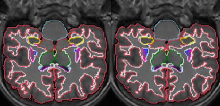
An example of a segmentation algorithm. On the left is the true markup; on the right is the obtained markup. The
authors of [ 1 ] managed to obtain the following segmentation accuracy for anatomical structures (the DSC indicator is a measure of Dyce's similarity):
- Cerebellum - 0.885 ± 0.05
- Pallidum - 0.7442 ± 0.009
- Ventricle - 0.9 ± 0.02
- Blood vessels - 0.2 ± 0.001
- Midbrain - 0.8474 ± 0.0073
Sources:
- A. Yu. Zubov, O. V. Senyukova. GraphiCon 2015. Image segmentation of magnetic resonance imaging of the brain using multi-atlas mapping
- Sequence-independent segmentation of magnetic resonance images
Ultrasound Image Analysis
For example, let's take the task of finding the nervous structure of the neck from recent kaggle competitions .
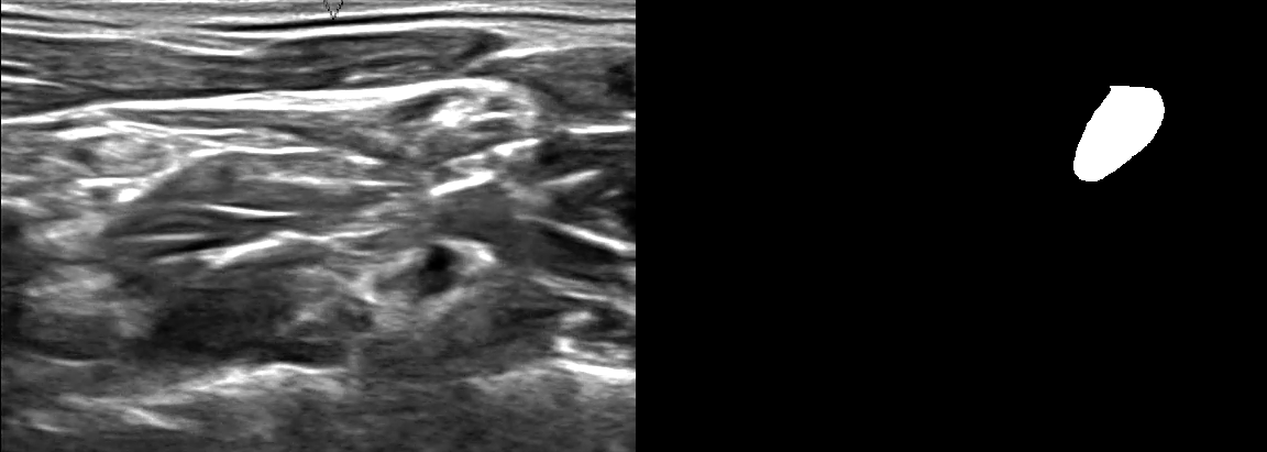
On the left is the ultrasound image of the neck, on the right is the nervous structure; the images show that finding the nervous structure is a non-trivial task even for a person (who is not a specialist in this field)
Accurate finding of the nervous structure in ultrasound images is an important step in the effective insertion of a catheter to block or reduce pain. Such catheters, in particular, help patients with drug addiction recover more quickly.
The best result, as Dice was similar to DSC on kaggle, was 0.73226.
Sources:
Speckle laser pattern analysis
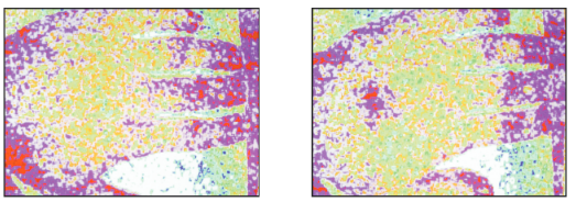
Laser speckle pattern of the hand, showing perfusion before and after rubbing a small area of the hand
Automatic analysis of laser speckle pattern is used to measure blood flow. In particular, it helps with laser therapy of damaged tissues.
Sources:
- Laser speckle contrast imaging for measuring blood flow
- Laser speckle contrast imaging in biomedical optics
Conclusion
The problems considered are only a drop of water in the sea. In the field of medical image analysis, there is great scope for research.
The rapid development of deep learning helps to gradually improve the accuracy of the developed systems for the analysis of medical images, which may soon be used everywhere. And this will undoubtedly increase the level of public health.