Medical students virtually operate on three-dimensional bodies
- Transfer

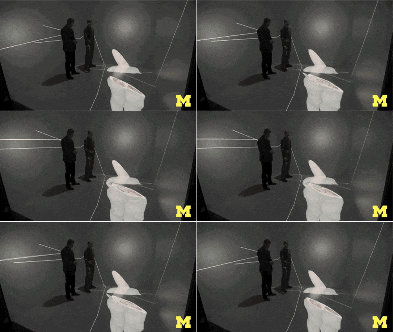
A three-dimensional image of the human body floats in the air, as if lying on an invisible cart. A few centimeters away is Sean Petty, an employee of the University of Michigan's 3D Imaging Laboratory. There are special glasses on Sean, and in the hands is a small joystick that allows you to arbitrarily “cut” layers from a three-dimensional cadavre, exposing tissues and organs. Sean zooms in and rotates the body for a better view of the details of the anatomy.

Assistant professor Alexander DaSilva believes that for him and his students such a chance fell once in a lifetime. “When I first saw this, I almost cried,” he says. Da Silva heads the Department of Head and Rotolacial Pain at the University Dental Clinic, as well as the Institute of Molecular and Behavioral Neuroscience Institute. “In my wildest dreams, I could not imagine that such a thing would be possible.”

The three-dimensional model allows virtual “showdowns” to be rolled back in case of unsuccessful actions. It is also possible to enlarge the image of body parts of interest. Detailed 3D-model at the moment allows you to "cut" very thin layers. The developers of the technology claim that it can be used in many other scientific disciplines, including technical and natural sciences. In particular, Alexandra DaSilva uses it to study the brain of patients suffering from migraine.
