How to interest a student in the natural sciences using a tomograph, molecular dynamics and augmented reality
Modern science education in most Russian schools serves one purpose: to discourage student interest in the subject. With nearly a decade of experience creating graphics for leading publishers of non-fiction, illustrations published in The New York Times and The Washington Post, and even images for presentations by Nobel laureates, we are determined to tackle this problem. This and the next post outlines the story of how we helped like-minded people from the laboratories of the Polytechnic Museumdevelop the format of the educational poster familiar to schools, providing students with an augmented reality application. In the process, we obtained a detailed three-dimensional model of the prepared frog based on computed tomography data, visualized the gene and its corresponding protein at the same scale, and also simulated an interactive atom that allows us to see the orbitals of different shapes and sizes.
Posters and posters are quite a familiar thing in the field of education, which has long been integrated into the educational process. Despite the abundance of digital whiteboards and other innovative equipment, posters are still used in schools - but often need to be seriously updated. Their obsolescence is a good reason for an enthusiastic teacher to replace an ancient poster with a more relevant one (also containing a surprise). We have no illusions that it is possible to solve the problem of motivating and involving students with just a poster, but a pilot project in which the poster is a “window” for accessing expanded content in a tablet or smartphone environment familiar to the student seems interesting to us to assess the potential of such ventures.
A flat illustration may well be informative and attractive, however, in a two-dimensional picture, it is not easy to really thoroughly and clearly demonstrate the structure of complex objects, which are quite a lot even in the school curriculum (from the structure of molecules or living beings to experimental facilities or atomic orbitals). I would like to look at a complex object from different sides in order to remove layer by layer or to see individual parts. This becomes possible if we combine an educational poster and a mobile application created using augmented reality technology, which we did. In addition, the application also contains “stripped-down” electronic versions of posters that adapt the content of the poster to a mobile device.
The topics of the first posters that teachers who visited the educational laboratories of the Polytechnical Museum will receive are “ Amphibian Anatomy ”, “ Atom Structure ”, “ DNA, RNA and Protein ”, as well as “ Solubility Table ”. It took us about four months to complete our work - the poster with the frog anatomy required the most comprehensive approach.
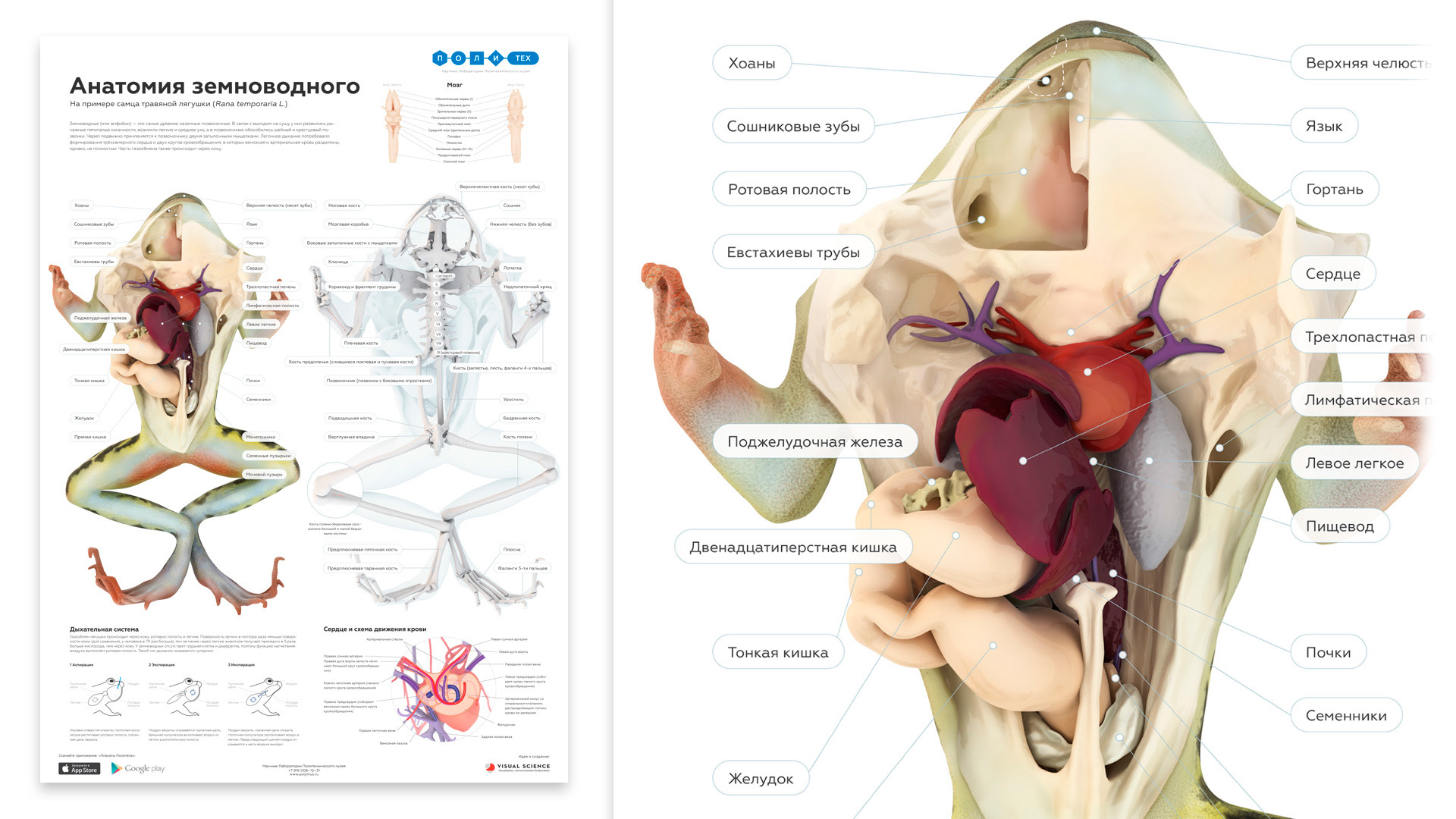
Frog anatomy
On the one hand, this topic has been developed for many decades: there are a lot of books, illustrations and videos showing how the frog works; schemes of individual organ systems and, of course, posters are available.
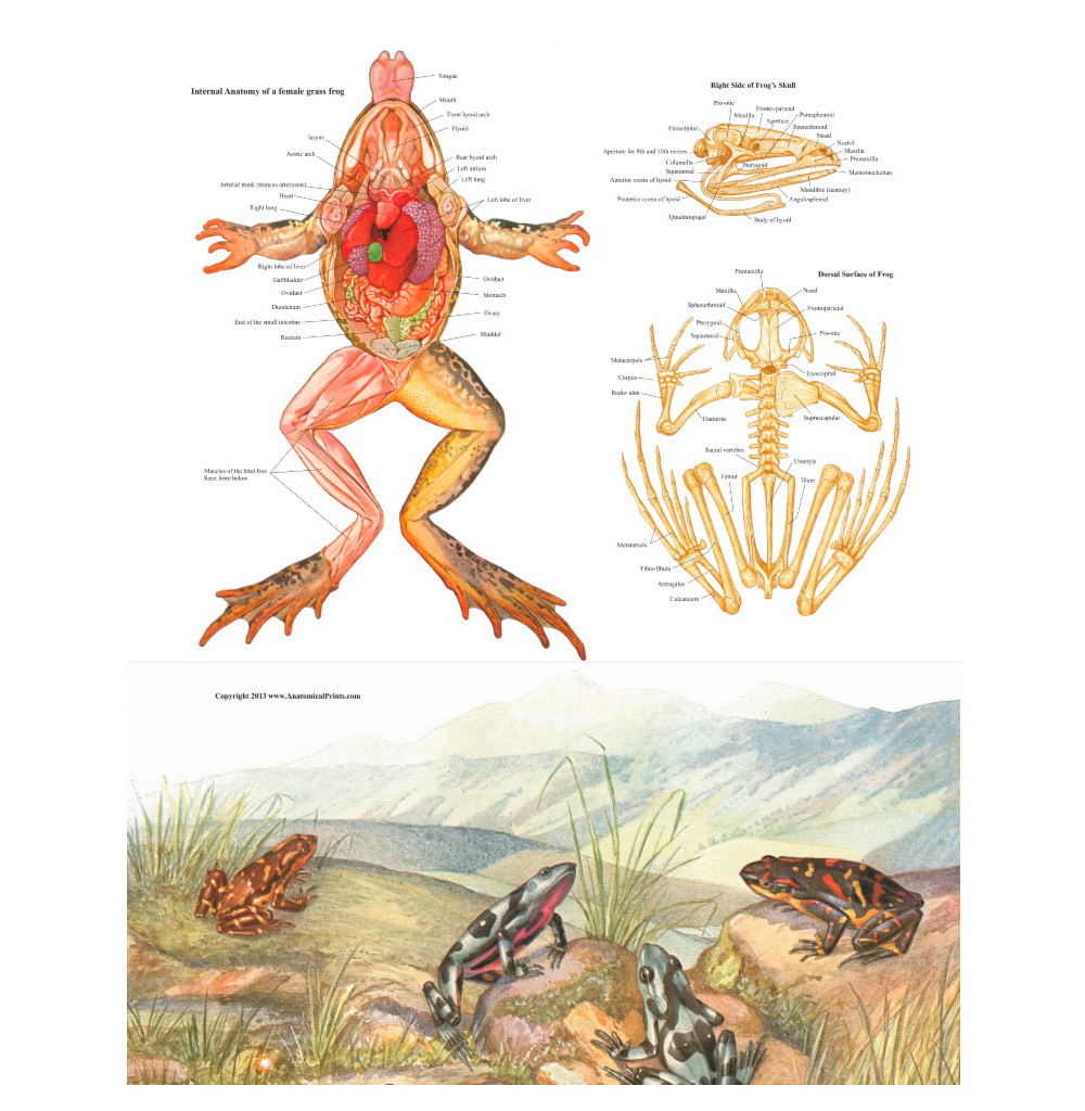
The picture is from here .
Existing illustrations are mainly made in the style of a classic monochrome design. This allows you to schematize the material, which is often good, but in most cases, these illustrations contain many inaccuracies and incorrectly reflect reality. Surprisingly, the most comprehensive Russian-language guide to the anatomy of a grass frog ( P. Terentyev’s book “The Frog” from the series “Laboratory Animals”) is illustrated very poorly and in fact contains many not the most obvious points, because of which there is almost no chance to understand the topic without zoologists involved in vertebrates. When it comes to the relative position of the moving parts of the body or, for example, the vessels relative to the skeleton, the animal should be dissected to understand the issue.
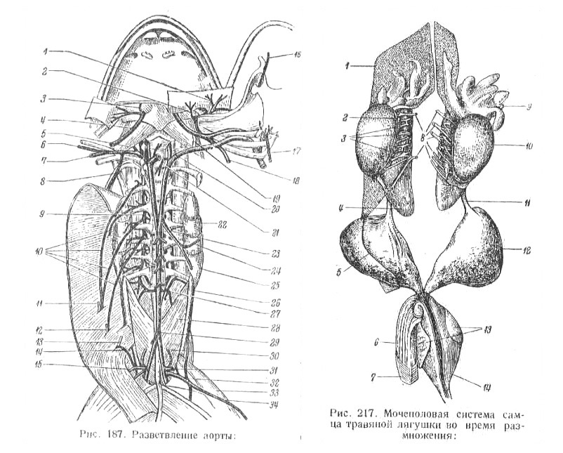
In the video preparation has its own problems: they can be shown not all that is required (most complex in this regard is the circulatory and nervous systems).
The second key problem of existing school textbooks is that they cannot attract and retain the attention of a schoolchild who is tempted by the work of the best specialists in the gaming, advertising, and film industries, who manage disparate budgets when visualizing even simpler objects and phenomena.
Our task in this case was to create the most accurate and close to reality manual that would be convenient to use to familiarize yourself with the structure of all systems of frog organs. And, of course, we wanted to “repel the smell of naphthalene” from biology as a discipline - at least in this particular case. Traditionally, we use hybrid approaches based on three-dimensional graphics. We had no illusions and plans regarding the fact that we would be able to draw and model a frog according to the existing atlases and illustrations - this approach obviously looked like a dead end. Therefore, we set ourselves the task of obtaining the most accurate tomographic scans of a frog.
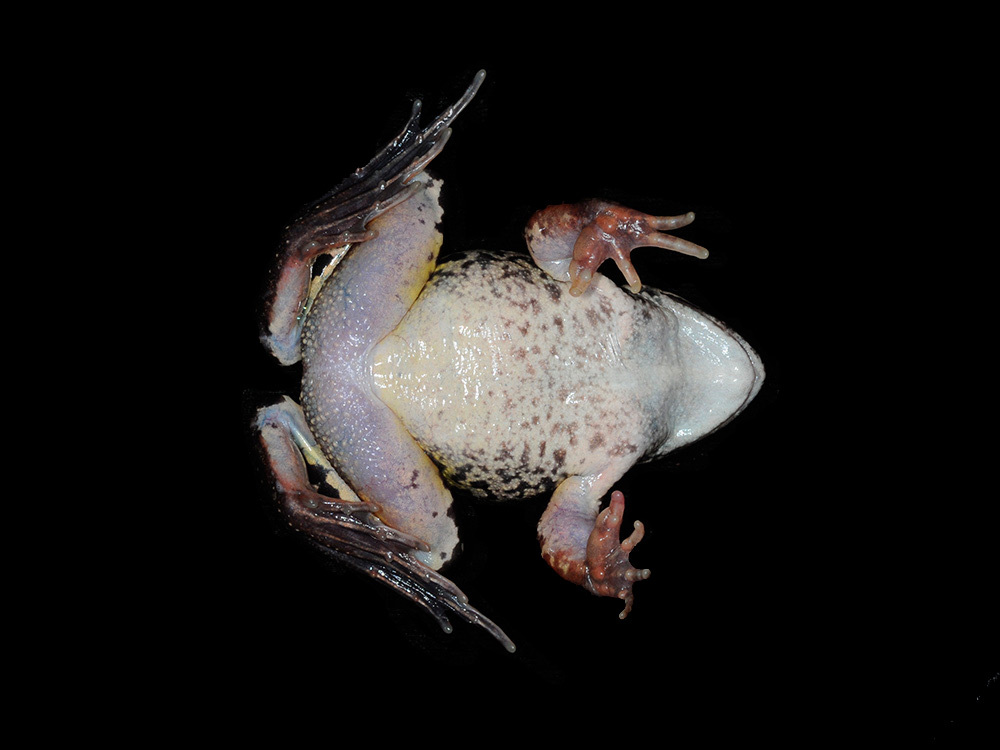
The search for the frog itself was not easy. Firstly, it turned out that by the end of May, when we needed animals, they had already ended the breeding season, and we could not find them in the forests near Moscow. In addition, zoologists and physiologists, who annually train students on frogs, note a sharp decrease in their number in the forests near Moscow, because of which frog hunters collect animals farther from the city. A search at pet stores and bird markets also did not return results - for approximately the same seasonal reasons. I had to look for people who are directly involved in catching frogs for terrariums, shops and educational institutions. As a result, we got three desirable representatives of the species Rana temporaria in a chilled and sleepy state.
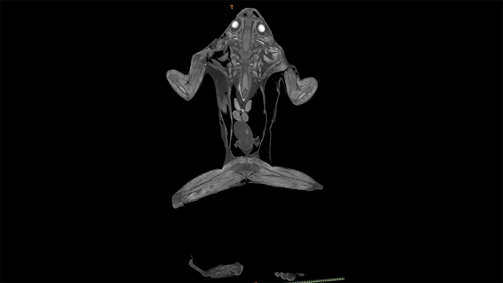
Modern methods of tomography make it possible to obtain a scan with a resolution of up to 20 microns (a micron is one ten thousandth of a centimeter). This means that about an eight-centimeter frog will have about 4000 horizontal “slices”. In our case, the layers were somewhat smaller, but still their number was more than sufficient to build a truly reliable model, where, for example, the phalanx of the animal’s finger and even rather small vessels can be distinguished. Only this approach guarantees the accuracy of the relative position of all organs and organ systems in the final model. Resolutions suitable for resolution are used to evaluate the magnitude of defects - for example, in electronics.
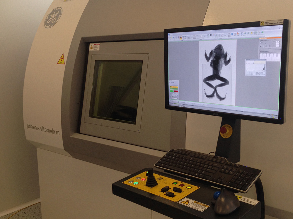
We managed to access one of these X-ray tomographs. It would seem that after that everything should be quite simple: we place the animal in the device, we get a scan. In fact, the problem of contrasting internal organs arises: in other words, the “tomograph” allows you to distinguish only fairly dense tissues, like bone, and the soft tissues in the images merge into a single whole. This problem is quite widespread, in connection with which researchers have already worked out various drug treatment techniques based on differences in the ability of different tissues to transmit and accumulate contrasting agents (usually substances containing atoms of relatively heavy elements, from halogens to heavy metals, play this role) . We had to thoroughly study the methodological scientific literature, and then turn to colleagues from the Faculty of Chemistry of Moscow State University for help in preparing the necessary reagents. But in this case, some tasks remained unresolved. In particular, bones, muscle tissue, cavities and organs after treatment become quite contrasting, but the circulatory system does not. As a result of the work, we had to adapt the existing protocols and develop new technology that solves this problem. At this stage, zoologists from the Faculty of Biology of Moscow State University joined our team, who helped dissect the animal and inject an alternative contrast agent into the circulatory system before tomography. cavities and organs after treatment become quite contrasting, but the circulatory system does not. As a result of the work, we had to adapt the existing protocols and develop new technology that solves this problem. At this stage, zoologists from the Faculty of Biology of Moscow State University joined our team, who helped dissect the animal and inject an alternative contrast agent into the circulatory system before tomography. cavities and organs after treatment become quite contrasting, but the circulatory system does not. As a result of the work, we had to adapt the existing protocols and develop new technology that solves this problem. At this stage, zoologists from the Faculty of Biology of Moscow State University joined our team, who helped dissect the animal and inject an alternative contrast agent into the circulatory system before tomography.

As a result, combining software for the analysis of computer tomograms with common programs for three-dimensional modeling, we were able to obtain a complete model of the frog along with models of all its main internal organs, blood vessels and skeleton. It took about a month of painstaking work on marking, “cleaning” and additional 3D-modeling.
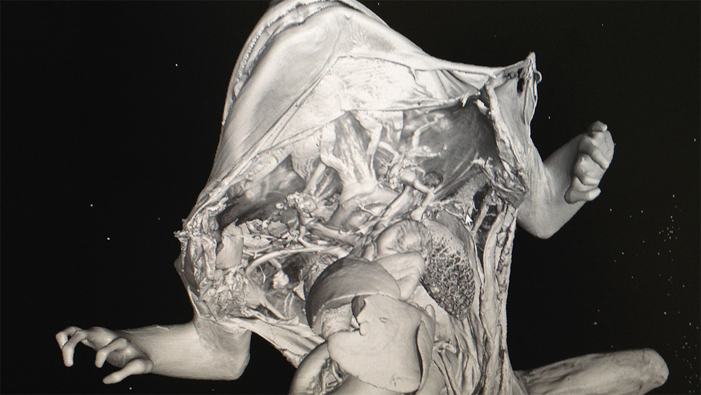
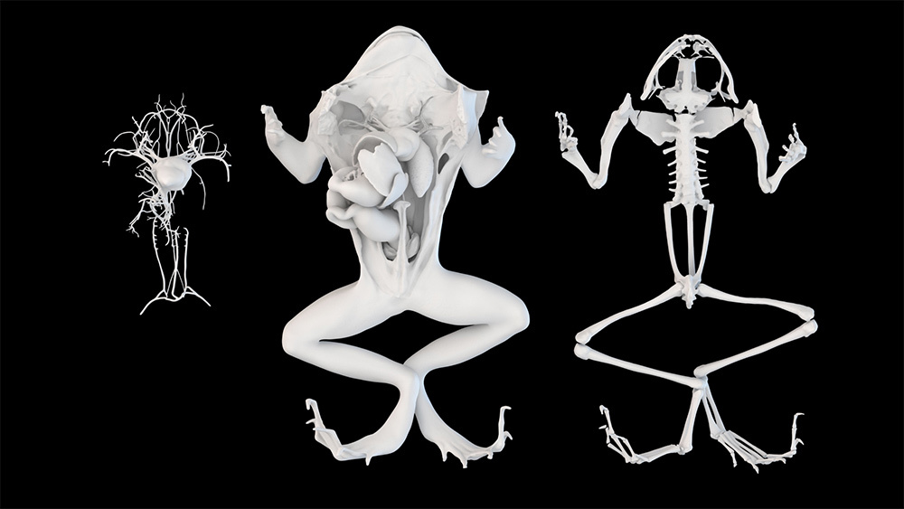
The images on the poster were created precisely on the basis of the obtained model, and in a special application with augmented reality for modern devices based on iOS and Android , it is possible to look at the model from different sides to see those structures that are not visible in the static illustration.
Such a comprehensive approach for a school poster may seem redundant. But, firstly, on the example of three-dimensional models of viruses, which were not created for schools at all, we noticed that schoolchildren appreciate it when they are treated in the same way as older people. They like it when they are offered together to sort out something at first glance complicated, inviting them to rise to the level of a scientist, instead of looking at schemes of unknown artists with template definitions from a textbook. Secondly, we have no illusions that all textbooks should be illustrated in this way, but we believe that in school education there is a place for bright and interesting “entry points” into the subject, where we can make every effort and still captivate schoolboy with biology, chemistry or physics. Thirdly, anticipating the questions of green and zoodefenders, we want to convey the idea
In the next post, we will talk about creating an Atom interactive poster and an insulin gene model.