Scientists for the first time compiled a 3D model of the Drosophila brain
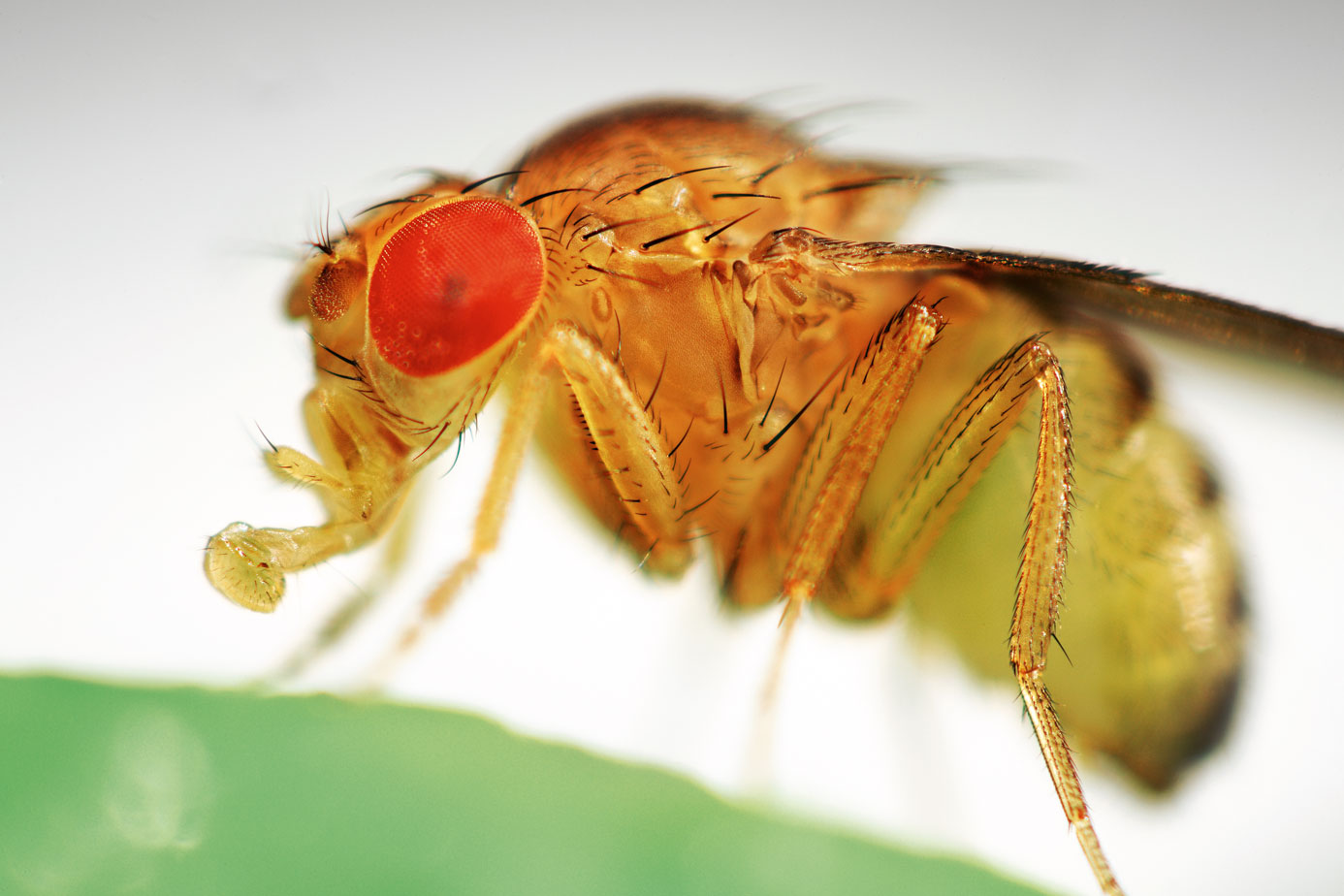
Black-bellied Drosophila (source: geo.ru) Scientists have been
studying the nervous system of humans and animals for hundreds of years. Of course, during this time a person began to understand much better the principle of operation of individual nerve cells and the whole system of which it consists. But to a complete understanding is still far.
The study is conducted according to the principle “from simple to complex”: if there is no possibility to immediately understand how, for example, the human brain works, then experts are studying the brain of simpler creatures. Scientists from the University of Tokai have chosen as an object for studying the brain Drosophila.
The brain of even such a small insect as a fruit fly is a very complex system. It took a long time for scientists to build a three-dimensional model of this body. Now experts are working with a number of techniques that allow you to study the structure of the brain without any problems. For example, scientists use fluorescent substances that highlight individual neurons. The electron microscope also helps in studying the brain, showing its structure at the neural level.
After individual neurons are “mapped,” the resulting images are analyzed to create a single system. The mapping of connections between neurons and the construction of a brain model is the ultimate goal of such work. All this is necessary in order to understand how everything is connected with everything, and how it all works.
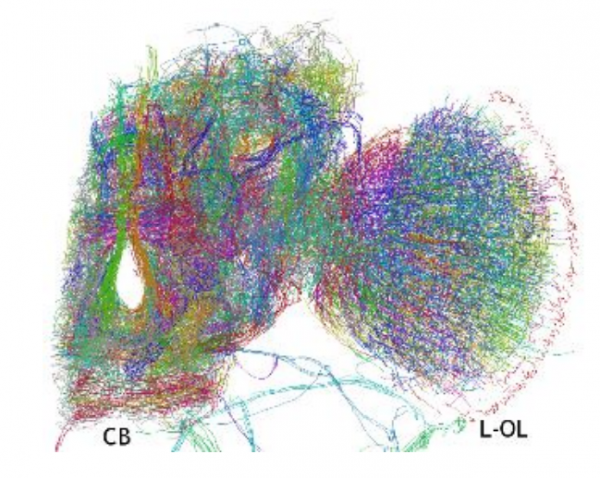
The three-dimensional model of the brain, created using a computer, demonstrates many neural connections. A team of scientists from the University of Tokai, under the leadership of Ruta Mizutnani, has developed a new method for drawing up a three-dimensional map of the brain. For this purpose, a special substance is used, the individual molecules of which are attached to the neurons of the brain. Next, scientists create a "skeleton map" of molecules by irradiating the brain with x-rays. Using their method, scientists were able to make a detailed three-dimensional map of the network of insect neurons in the brain.
In biochemistry, a special method is used to create 3D models of complex organic substances. To create a "skeleton map" of the compound molecule, X-ray radiation is used. If possible, the compound of interest is crystallized. And further, X-ray crystallography is used (X-ray structural analysis). This is the use of X-rays to identify the molecular structure of a crystal. The method is based on the phenomenon of X-ray diffraction - X-ray beam scattering by the atomic structure of a crystal.
This method is not bad, but if the structure of a substance is very complex, then it takes a lot of time to compose a 3D model of a substance molecule. For the past couple of decades, computers have been assisted by scientists who analyze the data obtained during the study of a substance. Computer systems help to assess the position of an atom in three-dimensional space, then the relationship with another atom is studied, then with one more, and so on. Software gradually builds a model of the substance under study.
 Mizutani decided to use this method and software to determine the location and shape of the neurons of the Drosophila brain. There are certain difficulties here, one of which is that neurons are not atoms at all. These are complex objects that can sell connected to each other in the most unusual way.
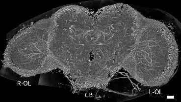
To construct a map of the brain, scientists used a method called x-ray tomography. This is a method for layer-by-layer studies of the structure of inhomogeneous objects in X-rays, based on the dependence of the linear absorption coefficient in the X-ray range on the composition and density of the substance. Scientists infiltrated the brain of the fruit fly with silver paint, and then illuminated with X-rays. A special system helped assess the deviation of X-rays. And this, in turn, made it possible to create a three-dimensional map of dye molecules absorbed by neurons.
After that, scientists moved to the next stage of work, using these data to assess the location and shape of brain neurons. In the course of work on the creation of a Drosophila brain map, special software was used. It allowed to make sure that the system would not regard two adjacent neurons as one, for example. The software gradually mapped the front sight of the brain, looking for anomalous data and checking them for errors. The result of the software was checked by the operator - a man. If something was wrong, the person corrected the problem.
The final model shows100,000 neurons. The system tracked 15,000 links between them. To create the map, according to scientists, it took about 1,700 man-hours. But the result was worth all the effort and time spent. 3D-model of the brain Drosophila is the first in the world. With its help, it was possible to identify already known formations in the brain of an insect, as well as to detect structures about which scientists knew nothing.
The work of the Japanese is very important for the further study of the nervous system of animals and humans. Over time, scientists hope to create a map of the brain of more complex organisms.
Drosophila due to a number of its features are a popular object for study. A year earlier, the team of scientists from the Howard Hughes Medical Institute in the USA presented an interesting video showing clearly the nervous activity of the fruit fly larvae.
Scientists studied the nervous system of the larvae while moving back and forth. The video shows a noticeable signal transmission from the upper part of the larva to the lower body and back. According to experts, this model is very detailed. She managed to create through the use of new methods of shooting the neural activity of the body.
To make all this possible, Philipp Keller ( by Philipp Keller ) and Misha Ahrens ( Misha Ahrens ) genetically modified fruit fly. The modification was to make each neuron of the nervous system of this organism fluoresce when receiving or transmitting a signal. When moving, an optical system was used, which allowed the flies to take pictures simultaneously from both sides.
The scientific work “Three-dimensional network of Drosophila brain hemisphere” was published on September 8, 2016 (DOI: 10.1016 / j.jsb. 2013.08.012).