"Invisible" flies: a new method of studying the nervous system through tissue depigmentation
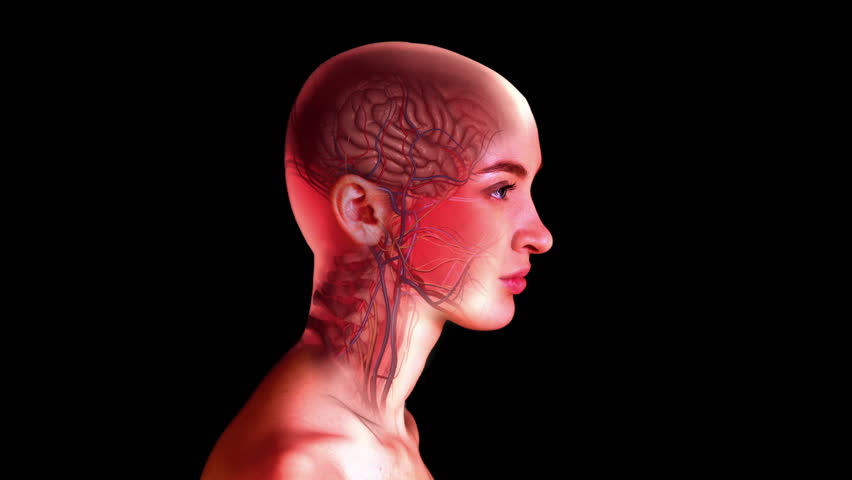
The study of any organism is a complex and demanding process. Modern scanning methods have succeeded in this. Today we can study the body or a separate organ in detail in three-dimensional images. About 100 years ago, a three-dimensional view of organs could only be obtained by removing them from the body and examining them live, so to speak. However, there is always some "but." But even the most accurate scanners and microscopes can not give 100% accuracy. Now, if it were possible to make everything unnecessary invisible, and leave that part of the body that we want to study visible. Sounds like science fiction, doesn't it? I agree. But now it is real. Today we will get acquainted with the study of a new method of scanning organisms on the example of Drosophila. How do scientists manage to make an ordinary fruit fly "invisible" How accurate is their scanning method and how will it help in the diagnosis of human diseases? We can get answers to these and other questions only in the research report. So let's not pull. Go.
Basis of research
As I said in one of the previous articles , the main character of which, like today, was the fruit flies, this small and, in truth, annoying insect is the subject of much research. With its help, scientists are studying quite a lot of things: from aerodynamics to the nervous system. And it is about the latter will be discussed today. The study of such complex systems in such small organisms is associated with certain difficulties. We are already familiar with the methods of studying organisms by cutting them into many ultra-thin layers. This method makes it possible to study many systems in more detail. However, regarding the nervous system, this method, according to the researchers, violates cellular integrity, which complicates the process of researching individual parts of the system.
If we apply scanning on small organisms without physical “lamination”, we will see all the systems at the same time. But scientists want to explore only one system, while others should not interfere with this process. Therefore, a brilliant and simple idea came to them - to make everything invisible (more precisely, transparent), and leave the desired system intact.
At the moment there are enough tools for the implementation of such an idea, it remains only to combine them together. The main ingredients of the new scanning method called FlyClear (in honor of the first test subject - Drosophila flies) are tissue “cleaning” and ultramicroscopy.
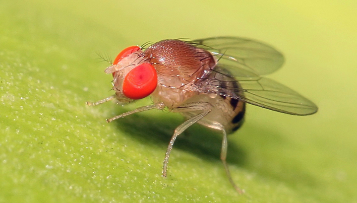
Drosophila was chosen as a test subject for a reason. The basis of most tissue cleaning methods is the genetic modification of the test organism. In this case, it is fluorescence. This process is extremely complex and time consuming. The larger the body, the more time it takes to modify it. In addition, not all creatures can be modified as scientists want. Drosophila, on the other hand, gives in to such genetic manipulations quite easily, and this process takes not so much time.
At the moment, the most popular method for the study of organisms is confocal microscopy, but it is produced only after dissecting the tissue. The question arises - and if not dissect? In this case, we get a low-quality pale image, and the process will take a lot of time. There will also be strong absorption and scattering of photons in the sample, especially with increased tissue pigmentation (as in Drosophila). Accordingly, the sample must be cleaned, that is, made transparent. Thereby to neutralize the decrease in signal intensity and to achieve uniformity of spatial resolution. The main way of "flipping" (sorry for the made-up word) of the tissues is to reduce the refractive index at the boundaries between the cellular components.
Researchers do not deny that now there are several ways that have already become classic, to make the fabric transparent. However, they also claim that there are a number of drawbacks with these methods. The preservation of morphology, the stability of fluorescence, the depth of scanning, and so on - with all these aspects of existing methods, to put it mildly, do not fit. In addition, none of them gives a complete depigmentation of tissues.
It turns out that it is necessary to carry out a cross-section of tissues for better scanning of small bodies? Not really. Scientists cite as an example all the same fruit flies. Located in its peripheral regions (legs, eyes, antennae), the Drosophila sensory neurons have very long connections with numerous nerves and the brain. If we examine these regions in terms of clarification, we will not get a complete picture of how the nervous system works in these regions.
In this study, the FlyClear method allowed us to remove the pigmentation of Drosophila tissues, exposing its nervous system. It took about a month. And ultramicroscopy allowed us to visualize the smallest neural connections and build a complete map of the midge's nervous system. Also, a large role was played by the substance, with the help of which it turned out to “stain” the nervous system in a bright green color - green fluorescent protein (hereinafter referred to as GFS). The gene of this protein, derived from the jellyfish Aequorea victoria, is used specifically for gene modification of the test samples.
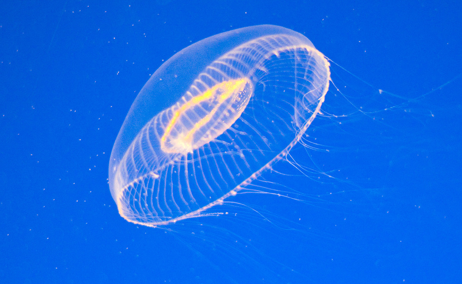
Medusa Aequorea victoria.
The data obtained by fluorescence microscopy, were processed by a special algorithm that allows you to create a three-dimensional image with high resolution.
Preparation for the study
Optical cleaning of Drosophila tissue is not an easy process. The chitin exoskeleton and photopigments of the faceted eyes are the most complex regions of the midge's body for the procedure. Currently available methods of tissue cleaning are far from ideal, because the researchers have created their own - FlyClear, I mentioned about it earlier.
FlyClear combines several previous methods. The first of these in the process is CUBIC (clear, unobstructed brain imaging cocktails). Further steps depend on the state of the sample (larva or adult).
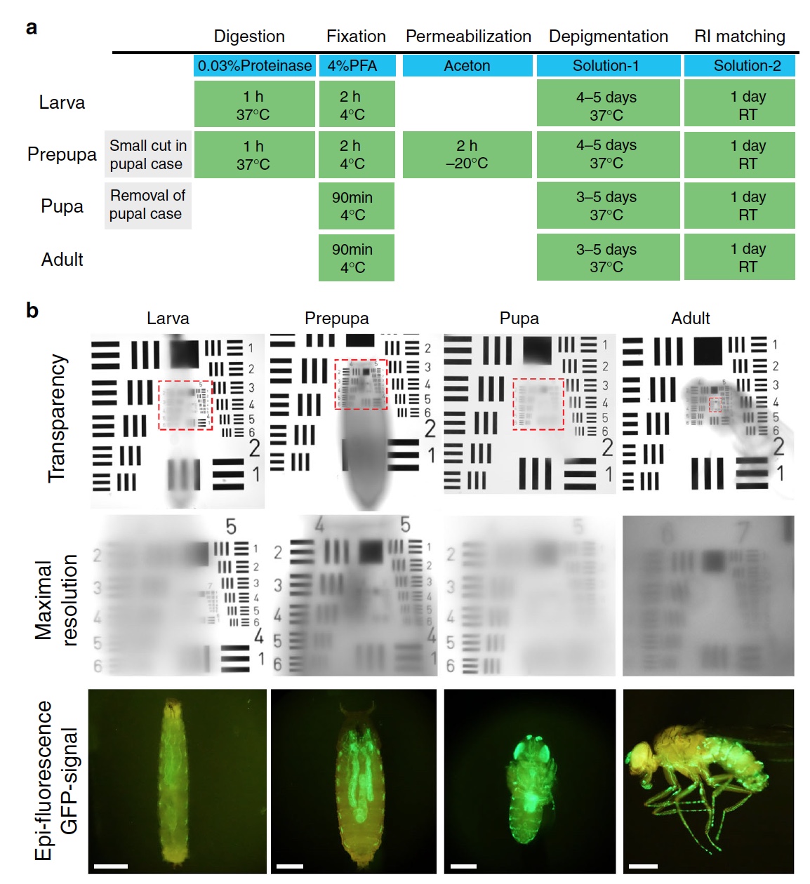
Variants of the FlyClear procedure.
In the case of the larva, 0.03% protease and formaldehyde fixation are used. The fact is that protease cleaves peptide bonds between amino acids of proteins, and this helps to "discolor" the tissue. Next, acetone is used on the sample to conduct permeabilization (changes in the permeability of the cell wall).
Scientists have been able to improve the stage of CUBIC, replacing [CH 3 CH (OH) CH 2 ] 2 NCH 2 CH 2 N [CH 2 CH (OH) CH 3 ] 2 (N, N, N ', N'-tetrakis (2-hydroxypropyl a) ethylenediamine) on C 10 H 24 N 2 O 4(2,2 ', 2 ", 2"' - (ethylene dinitrile) -tetraethanol)). This change allowed to achieve complete depigmentation of tissues, including in difficult areas (eyes and epidermis).
So, the complete tissue depigmentation method works. Now you need to check the second important stage of the study - fluorescence. More precisely, it is necessary to check whether the new reagent C 10 H 24 N 2 O 4 will suppress the fluorescence signals, given that Drosophila already have low fluorescence after genetic manipulations. For the test, the samples (Drosophila bodies) were divided in half: one half was without treatment, the second with processing. The analysis showed that the level of severity of GFB and the truth decreases when using C 10 H 24N 2 O 4 , however, this does not affect the overall accuracy. And all due to the fact that the same reagent allowed to achieve a high degree of tissue depigmentation. That is, the advantages of the reagent leveled its shortcomings.
The finished sample should be considered, so to speak, and for this purpose, advanced ultramicroscopy was used, which allows to obtain an image of the sample with the same resolution in all planes.
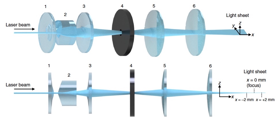
Optical system: 1 and 3 - a plane-convex aspherical cylindrical lens; 2 - Powell lens; 4 - elliptical apodizing soft aperture; 5 and 6 - acylindrical lenses.
Typically, standard ultramicroscopy uses cylindrical lenses and a rectangular aperture. In this study, additional components were used that change the shape of the beam to an ultra-thin one with improved characteristics.
The use of spherical lenses due to the ability to obtain an image without distortion and with minimal aberration.
The standard aperture has also been changed to an elliptical apodizing soft aperture (in the image above number 4). The new aperture allowed to eliminate unwanted intensity distribution in the optical system.
Research results
Below are the specific systems of the Drosophila organism (respiratory, visual, nervous, etc.) illuminated by the ZFB, while the rest of the tissues are completely depigmented.
In order not to stretch the article, he hid everything under the spoiler.
The third stage of the larva: trachea, digestive system and salivary glands.
Predictation: the developing visual system and the innervation of the segmental nerves into the abdominal nerve circuit.
Extremities of an adult Drosophila.
Spinal ganglion.
Dolly: visual and olfactory system.
The sensory neurons of the eyes, antennae, maxillae (the second pair of jaws) and labellum, as well as their connection with the central nervous system of an adult.
Relationship of the antennal nerve and the antennal lobe in the adult.
Adult Drosophila.
Scientists without hesitation call the greatest achievement of their research the ability to create a full-fledged three-dimensional map of the nervous system. And if we consider that this system belongs to such a small organism (Drosophila), then such an achievement becomes even more significant.
The new technique allows you to accurately determine where and how certain neurons connect, both among themselves and with the brain. If a cross section were used, this would not have been achieved.
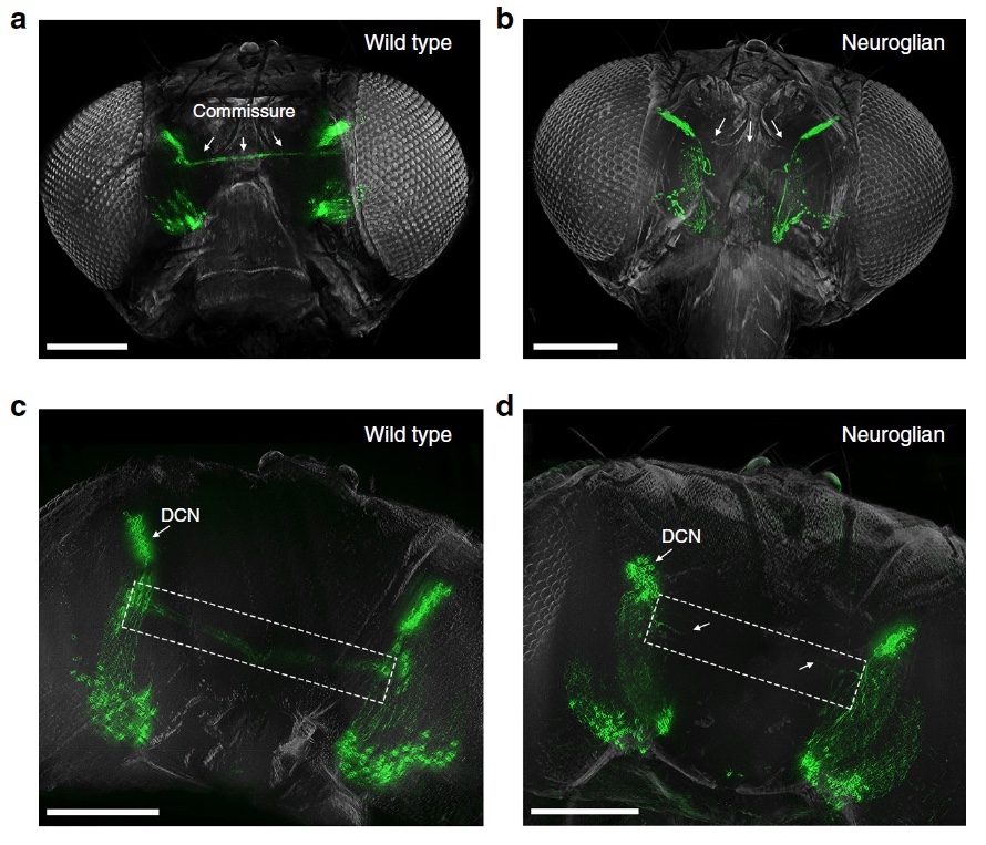
As a demonstration of the possibilities of their method, scientists show us two types of neurons of the Drosophila visual system: DCN - dorsal cluster neurons and MCN- cerebral columnar neurons (cortex columns in the posterior part of the brain). In the dorsolateral region of the brain, DCN clusters form commissural connections to excite the synaptic neurons of the medulla and lobula in the optical lobe. (images above a , c ).
Images b and d show a loss of commissural connection in homozygous mutants of Neuroglian neural cells. These changes in the nervous system are associated specifically with the FlyClear cell cleaning process.
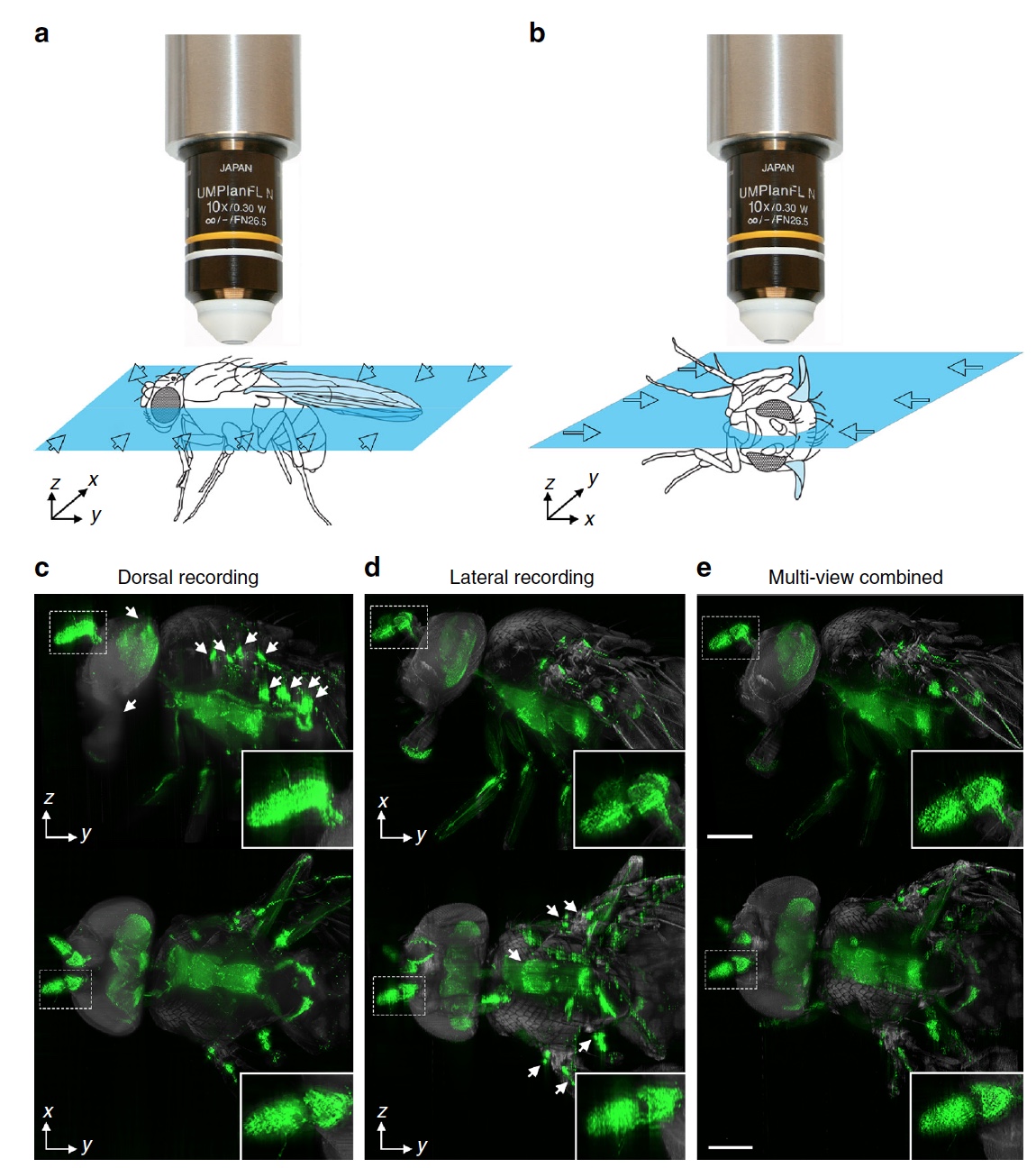
To construct a three-dimensional reconstruction, pictures were taken from two orthogonal directions, which were later merged. The algorithm based on 3D FFT determines the details of the sample that look clearer in one of two stacks of images. Further, it all came together in one three-dimensional reconstruction.
Those interested in more detailed study of the study can read the report of scientists and additional materials to it.
Epilogue
Scientists are not in vain boast and are proud of their work, because this was not previously. The method of lamination by cross-section was dominant in the field of research of organisms. But this method, although it has many advantages, has huge drawbacks. In particular, the cuts violate cellular and tissue integrity, thereby complicating the process of reconstruction of a particular body system.
To explore an organism with a complex nervous system, when every detail is important, will now be much easier. Scientists will be able to obtain more data, which will allow them to describe these or other processes in the body in more detail.
Also, do not forget that Drosophila is only experimental in this study, and not its basis. Improving the new scan method can serve as a person. And not only to study the body and its constituent elements, but also to diagnose diseases that can hide in the early stages where currently available scanning methods cannot identify them.
Research reveals many new things: new types of animals, new substances, processes and phenomena. But without research aimed at creating research tools, all this would be impossible.
And, of course, Friday offtop:
Сегодня в свете софитов опять были мухи. А где мухи, там и пауки. Но далеко не все пауки страшные на вид. Некоторые из них даже весьма неплохо танцуют.
Надеюсь это видео заставило вас улыбнуться (и может даже начать чуть меньше боятся пауков).
Спасибо за внимание и хороших вам выходных, ребята.
Надеюсь это видео заставило вас улыбнуться (и может даже начать чуть меньше боятся пауков).
Спасибо за внимание и хороших вам выходных, ребята.
Thank you for staying with us. Do you like our articles? Want to see more interesting materials? Support us by placing an order or recommending to friends, 30% discount for Habr users on a unique analogue of the entry-level servers that we invented for you: The whole truth about VPS (KVM) E5-2650 v4 (6 Cores) 10GB DDR4 240GB SSD 1Gbps from $ 20 or how to share the server? (Options are available with RAID1 and RAID10, up to 24 cores and up to 40GB DDR4).
VPS (KVM) E5-2650 v4 (6 Cores) 10GB DDR4 240GB SSD 1Gbps until January 1 for free if you pay for a period of six months, you can order here .
Dell R730xd 2 times cheaper? Only we have 2 x Intel Dodeca-Core Xeon E5-2650v4 128GB DDR4 6x480GB SSD 1Gbps 100 TV from $ 249in the Netherlands and the USA! Read about How to build an infrastructure building. class c using servers Dell R730xd E5-2650 v4 worth 9000 euros for a penny?