Scientific, medical illustration and animation: how doctors and scientists communicate with each other, students and the rest of humanity

With this post we want to give a general idea of the sphere in which we work. In the USA, Europe, Canada and Australia, the public is aware of it, since the field of scientific and medical illustration itself is developed much better than ours. We will recall the history of the issue, describe how things are now, what tasks are solved by the main biomedical imaging studios, tell about organizations that research and supervise this market, and share some statistics.
What is this all about? From drawing corpses to three-dimensional animation of molecules and viruses
In our body there are about 206 bones of various shapes and sizes, more than 600 muscles and a difficult to count number of nerves, blood vessels, and even more so all kinds of cells related to various tissues and organs. If you delve deeper, a picture of a huge number of proteins, RNA and other molecules opens, each of which performs a certain function, and small violations can have a dramatic effect on the vital functions of the body. It is enough to recall prions, which, due to improper folding of the molecule, can cause serious diseases.
Living systems are so complex that it is impossible to describe them in detail using words or formulas. Nevertheless, the description is absolutely necessary for understanding the mechanisms of work and the reasons for the breakdown of certain biological structures. To solve descriptive problems, doctors and natural scientists from ancient times have learned to make correct sketches and diagrams of both the structure of our own body and the surrounding animals and plants that can benefit us or cause harm. If at the time of Galen, medical and scientific illustration was naive and not accurate, which, by the way, did not prevent his work from being in demand for more than 1300 years, then, starting with Andreas Vesalius , it began to take shape as a discipline and an important tool in the training and work of doctors and scientists .

Examples of early medical illustrations. ( Source ).

Parts of the human body depicted by Andreas Vesalius. Page from De Humani Corporis Fabrica, 1543 ( source ).
Illustration as a scientific method received a new incentive for development after the first magnifying devices began to appear, and Hook , Levenguk , Malpigi , Grue and other natural scientists began to work, who began to describe the microworld and prepared the basis for the emergence of cellular theory and understanding of the microscopic anatomy of living objects .
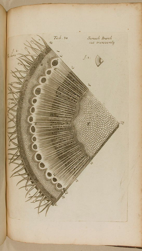
A cut of the stem of a plant. Illustration by Nehemiah Grew. From the book “The Anatomy of Plants. With an Idea of a Philosophical History of Plants, and Several Other Lectures, Read Before the Royal Society. ” 1682 year. ( Source )

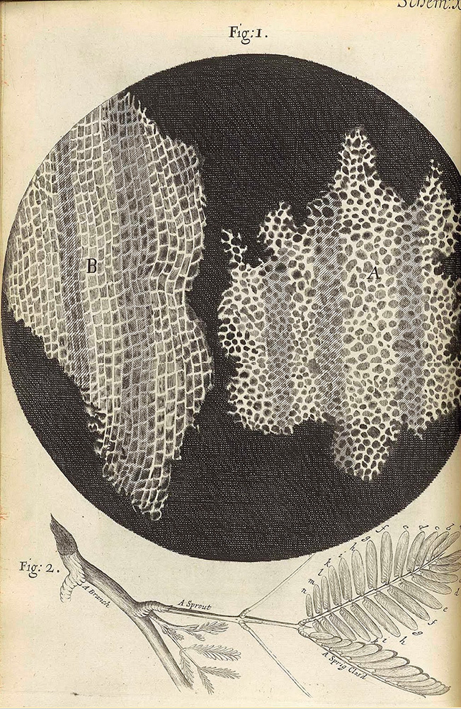
Robert Hooke's illustrations in the Micrographia book published in 1665 ( source ). Thanks to such drawings, living cells got their name and attracted the attention of researchers.
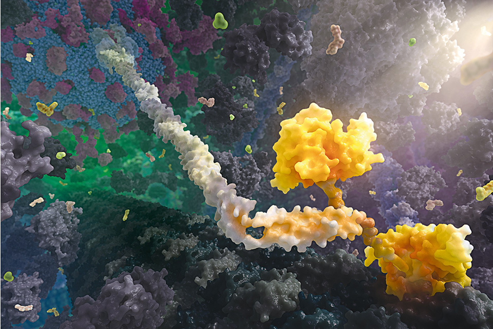
Currently, biology students in the learning process are still sketching microscopic preparations and cockroaches uncovered with a scalpel, however, scientific illustration has long been a separate integrated discipline that solves mainly communication and educational tasks, not research ones. The relevance of this type of activity is growing, since the volume and complexity of scientific and medical knowledge increases faster than the ability of people to understand it, and schemes, models and animations make it possible to study complex topics much more effectively. If in the sixteenth century a doctor and part-time medical illustrator walked through cemeteries at night in search of bodies for opening and sketching, now such radical approaches are not required, and a wide range of specialists is engaged in the field, from scientists to professionals in graphic design, three-dimensional modeling, computer animation. Of course, tremendous progress in science and scientific methods plays a major role in the evolution of medical illustration. Electron microscopy, X-ray diffraction analysis and molecular dynamics have shifted the attention of specialists in this field from anatomy and microbiology to molecular medicine, cell biology and biochemistry. Thanks to new methods, it became possible to identify and demonstrate X-ray structural analysis and molecular dynamics have shifted the attention of specialists in this field from anatomy and microbiology to molecular medicine, cell biology and biochemistry. Thanks to new methods, it became possible to identify and demonstrate X-ray structural analysis and molecular dynamics have shifted the attention of specialists in this field from anatomy and microbiology to molecular medicine, cell biology and biochemistry. Thanks to new methods, it became possible to identify and demonstratethe structure and mechanism of work of individual molecules , large molecular complexes, and even viruses .
A kinesin protein that moves a cell vesicle along a microtubule. Studio XVIVO . year 2013.
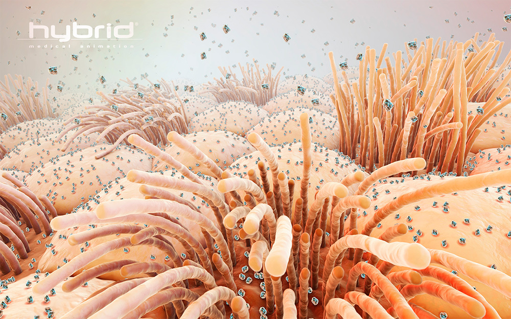
Particles of influenza virus on the surface of epithelial cells of the upper respiratory tract. Hybrid Studio .
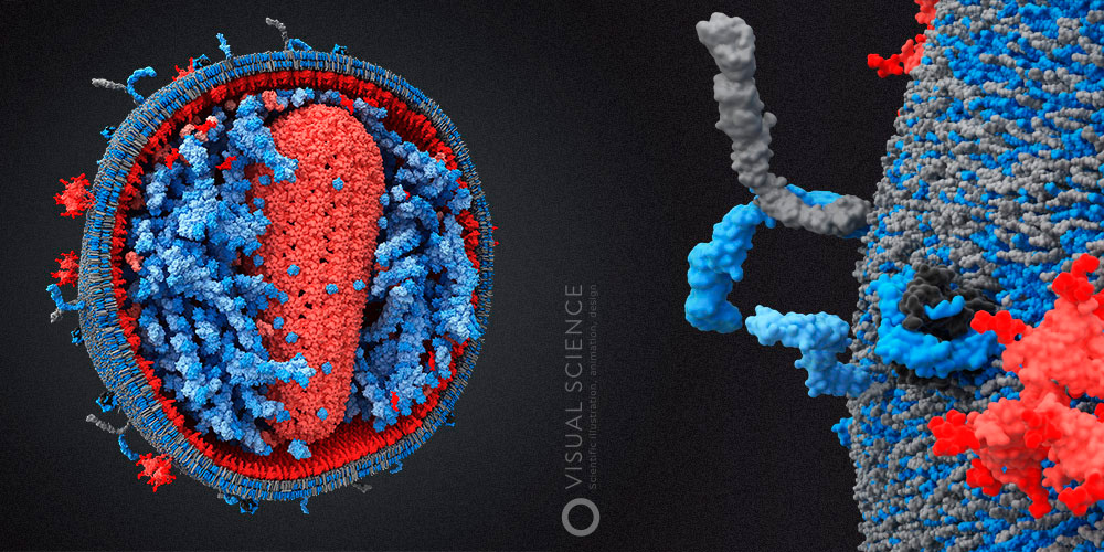
The internal structure of the human immunodeficiency virus . Visual Science Studio. 2010 year.
Who needs scientific and medical illustrators?
Many high-tech enterprises, whether they are farm companies, medical equipment companies, or private clinics, need to inform potential customers, partners, or investors about the essence of their developments and the services offered. Moreover, if they come to a design studio with a wide profile with an order for the production of presentation materials, a website or animated videos, most likely, obtaining an adequate result will be very time-consuming and long. The reason is that an ordinary designer or illustrator may not have the slightest idea about, for example, how the nanoparticles based on the copolymer D, L-lactide and glycolide are arranged, or whether Le Faure’s fracture affectsthe first type of ethmoid bone. Of course, you can go into lengthy explanations, throw designers with references from atlases and links to Wikipedia, but this approach seriously slows down the process and at high energy costs does not guarantee the desired result. Moreover, the time spent by a specialist on the client’s side, who “teaches” the designer, the number of edits and iterations, makes the total cost of this approach very high. That is why a specific niche of biomedical illustration, animation and design has formed, in which usually either individual specialists or teams combine competencies in the field of graphic design, medicine and science.
In addition to high-tech companies, high-tech design, of course, requires universities and other educational institutions and projects. According to the latest report of the American Association of Medical Illustrators , about 39% of specialists in this field work on a permanent basis at various universities and academic institutes. Well- known examples include David Godsell , who works at the Scripps Institute in California, the medical animation group at the Walter and Eliza Hall Medical Research Institute in Melbourne, and Graham Johnson , who develops imaging and modeling algorithms for molecules and cell structures at the University of California, San Francisco .


The internal structure of Mycoplasma mycoides. David Godsell. 2011.
These illustrations are very impressive in their detail. It is noteworthy that they are all made in pencil and watercolor. David outlines the contours of molecules in molecular imaging programs and then manually forms such an ornament. The disadvantage of this approach is that Godsell, as an artist, can interpret the forms of some objects in his own way, without delving into scientific data, which affects scientific credibility. Plus - that this way you can make many illustrations of even very large objects in a short time.
A little more than a third of medical illustrators have pharmaceutical companies, medical centers and clinics, publishing houses, law firms specializing in healthcare (clients often need to illustrate, for example, human injuries ), as well as advertising and design, as clients and employers. studios. The rest of the specialists surveyed by the association were employed in non-profit organizations and foundations (18%), and also worked for certain government offices (7%).
According to the study, in 2009 the average salary of medical illustrators was $ 61,000 per year, with a spread from $ 27,000 to $ 150,000. Studio managers and art directors earned more ($ 93,000 and $ 75,000 per year, respectively). Graphic designers, animators, and multimedia content developers earned approximately $ 47,000 to $ 58,000.
In total, the employment of more than 550 professional members of the association was analyzed.
Part of biomedical visualization is scientific illustration and animation. The specifics is the most accurate work with the source data, most often the results of experiments or in silico simulations. The tasks are the same - communication in a professional environment (starting with a certain level of fame of the author), presentation, illustration of scientific publications, textbooks and other educational materials.
Scientists in everyday tasks and on their own quite well find a common language with each other. Presentations at conferences are usually primitive in terms of graphic design and graphics, but in most cases they confidently solve their problem, because the audience is prepared and relatively undemanding to the level of presentation. Difficulties arise when a scientist has to explain something to an audience with a different level of immersion in the subject, such as students, or in general from another field, such as business, in the case of investors or partners at the stage of commercialization and popularization of developments. In this situation, it can be much more difficult to explain, and high-quality visual materials become necessary, so as not to be limited to the set of arguments of Richard Dawkins in this passage: coub.com/view/1j2dqvn .
Biomedical illustration now
Several large companies and quite a few smaller players and individual illustrators and designers are represented in the biomedical illustration and animation market. Most specialists in this field are concentrated in the United States, Canada, and very few in Europe and Australia. One of the leading animation studios, XVivo, has become widely known thanks to the large-scale three-dimensional video Inner Life of the Cell, which demonstrates the molecular processes that occur in the white blood cell when it is activated during inflammation. The video was made for Harvard's molecular and cellular biology education program, but quickly gained a much wider audience. When it was created, the company used the labor of Harvard students (rumor has it - for free).
Many well-known studios pay more attention not to intracellular processes, but to the anatomy and physiology of the human body. Examples of such work are demonstrated by Hybrid, Zygote , Nucleus , Brian Cristie , Invivo , Anatomyblue , AXS Studio . However, the matter is not limited to animation: the studios create educational three-dimensional models, illustrations and applications. This topic is relevant not only for educational institutions, but also for hospitals and clinics .
In order to get an idea of the field of scientific visualization, you can get acquainted with the works of participants in competitions that are held in this field. The most famous of them becameScience and Engineering Visualization Challenge , which is held by Science magazine in conjunction with the National Science Foundation, in which we also participated in 2010 and 2011. It emphasizes the natural sciences, and participants send both illustrations and infographics, as well as animations, and even educational games. Computer graphics and animation created in scientific design studios are also presented at SIGGRAPH contests , whose range of participants is not limited to medical illustrations. The effectiveness of communication and marketing in the field of medicine and healthcare is assessed by the jury of a competition organized by Medical Marketing & Media. Another noticeable, but somewhat losing ground competition is called Vesalius, and it focuses on the educational component.
The Association of Medical Illustrators, which has already been mentioned, is engaged in certification of specialists, coordinates some kind of union initiatives, conducts market research and produces analytics, and, by the way, has existed since 1946. They also hold their own competition and exhibition at the annual meeting of members, whose works can be found on the organization’s website. In competition, unlike SEVC, emphasis is placed on the medical component.

Photo from the first meeting of AMI. Philadelphia, Pennsylvania. 1946 year. ( Source )
In addition to competitions, a certain section of the region can givea catalog of medical illustrators , which is published annually, or photobanks specializing in this subject, where hundreds of thousands of illustrations are presented, from experimental photos to science art.
If the topic is interesting, in the following posts we will talk about a much wider field of communication in the field of biomedical areas of so-called healthcare communication, part of which is biomedical illustration and animation.
Only registered users can participate in the survey. Please come in.
Subject of the next post:
- 22.2% healthcare communication 54 business overview
- 14.4% Analysis and recommendations for scientific posters and illustrations from articles of readers and authors of Habr. Works can be sent to mailbox @. Let's make a post as typed 10-15 35
- 51.8% Continuation of the story about the process of creating 3D models of human viruses in Visual Science - part 2, molecular and 3D modeling 126
- 56.7% Medical anatomical illustration - the history of the study of the human body in the works of illustrators of 5 centuries 138
- 48.9% So-called science art or scientific art 119