MIS. Inserts and removed organs

In the IIA, the research protocol pattern is more like a constructor, which may consist of parts of different shapes and sizes. As a building material are the previously discussed tags and their attributes . With their help, you can add to the protocol all the fields that must be in it. However, sometimes there are cases when it is necessary to expand the capabilities of the current study protocol by adding additional measurement units to it. We called such additional parts inserts. In general, inserts can be an unlimited number. Templates of additional parts consist of the same tags as the template of the protocol itself, but other tags are used to indicate the insertion point and description of its type.
The description of the distant organs or anatomical parts of the body also requires special attention. They have nothing to do with the inserts, since the potential organs for removal are strictly in the standard study protocol. By default, the template contains all the anatomy described, and the absence is regulated by a special tag. It contains the status of the corresponding anatomy.
Let us consider in more detail the mechanics of the work of additional tags.
Inserts
One of the types of inserts - education . Its characteristic features are:
- the described object is initially absent in a healthy organism and appears over time due to some circumstances;
- the same type of the described object may be present in the body in more than one quantity.
Using the insert with the type of "education", you can describe the following types of pathologies:
- some pathological processes - a sequence of reactions that occurs due to the action of a pathogenic factor - a neoplasm (node, cyst, tumor), atheromatous plaques (contribute to the development of atherosclerosis), and so on;
- protrusions and hernias;
- stones (stones);
- wounds, etc.
If a doctor can familiarize himself with the dynamics of changes and comments for “standard” organs, then for formations in addition to the types of dynamics already mentioned, the dynamics of the development of education itself is also important. For this was added another entity - anatomy addon . In addition to viewing the dynamics, the preservation of each formation contributes to the description of the existing formations during the next visits of the patient, and not to duplicate the same ones.
Taking into account the fact that one type of described pathologies may occupy different organs or one organ in different places, such a concept as localization was introduced. Firstly, it allows you to clearly understand where the pathology is located or where exactly the measurement was taken. Secondly, it becomes possible to decompose the pathology only once in the “anatomy” and “measurement” entities, and not to create this pathology several times for each organ.
Consider a cyst. It is a pathological cavity in tissues or organs that has a wall and contains fluid. Its size, structure and content depends on many factors, and localization is one of them. According to the concentration of cysts within the same localization single and multiple cysts are isolated. Instead of creating anatomy of a single cyst of the left thyroid lobe, a single cyst of the right thyroid lobe, a single pancreatic cyst, etc., a single anatomy of a single cyst with the necessary measurements was added to the database. In turn, anatomy is already a localization: the left lobe of the thyroid gland, the right lobe of the thyroid gland, the pancreas, etc.
There is one anatomy in the human body, the status of which is not abated by disputes in various fields of science to this day. This is the fruit. For example, lawyers argue about the rights of an embryo, in bioethics they raise the question of recognition of the status of a person as the embryo and a fetus, etc. Within the framework of the IIA, we identified two important features for the anatomy of the fetus. First, this anatomy appears in the female body, and is not present there originally. Secondly, several fetuses can develop at once in the womb (multiple pregnancy). As the saying goes: “if something looks like a duck, swims like a duck and quacks like a duck, then this is probably what a duck is.” Taking into account the characteristic features of this anatomy, and not its essence, the fetus is added to the study protocol as the pathologies discussed above - with the help of the “education” insert.
The second type of inserts -additionally described anatomy , which we abbreviate as additional anatomy . In contrast to the objects described through the insert "education", the insert "additional anatomy" is intended for anatomy that are present in the body initially, but if they are not in an altered state, then they are not described. If the anatomy is changed, then it falls into the anatomy addon . Such anatomies include: the vertebrae of all parts of the spine, parathyroid gland, sublingual salivary glands, mammary glands, etc.
The third type of inserts is additional measurements . A few examples:
- in the study protocol of the examination by a urologist, measurements are added depending on the gender of the patient;
- in the protocol of the pediatrician's study, the inserts measure for children under one year old and the reflexes of children under one year old;
- a positive test for hepatitis C in the study "Anti-HCV antibodies (Anti-HCV)" test is performed to confirm the diagnosis of "viral hepatitis C", which checks for the presence of antibodies to some hepatitis C proteins;
- additional tests for the presence of salts and bacteria can be made in the analysis of urine;
- bacteriological sowing - microbiological laboratory testing of biological material by sowing it on certain nutrient media in order to detect the presence of pathogenic and conditionally pathogenic microorganisms in it - when confirming any bacteria using additional measurements, the sensitivity of this microorganism to a specific preparation is specified.
The inserts themselves are indicated in the template by the tags insertion-place and insertion-template-builder . A special button appears in the editable conclusion form on the place of the insertion-place tag . When you click on it, the doctor displays a dialogue with all possible formations, additional anatomy and additional measurements that are indicated in the template of this study protocol.
Insertion place
Used to indicate where to insert. Contains the insertion-template-builder tag.
Insertion-template-builder
Used to indicate a specific formation, additional anatomy or additional dimensions:
- id - number of the template in the database. Multiple values may be specified;
- anatomy-id - the number of anatomy in the database. Indicated for formations and additional anatomy;
- localization-id - the number of anatomy for localization in the database. Multiple values may be specified. Indicated for formations;
- name-overriden is the name of the insert. For formations and additional anatomy, the name of the anatomy is taken from the database by default;
- name-overriden-button - the names of the buttons in the insert selection dialog. The default value is from name-overriden;
- use-template-builder-name - flag to display the name of the template specified in the database. Options: true and false. Default is false;
- localization-marker-type - marker for the localization field. Options: none, word (spelled "Localization:"), icon (icon is inserted). The default is none;
- need-anatomy - a flag to display the anatomy field. Options: true and false. The default is true;
- need-name - flag to display an additional name field. Options: true and false. The default is true;
- need-status - flag to display the status field. Options: true and false. The default is true;
- need-localization - flag to display a drop-down list with localizations. Options: true and false. The default is true;
- need-found-date - flag to display the date field. Options: true and false. The default is true;
- need-template-builder — flag to display a drop-down list with template views. Options: true and false. The default is true;
- anatomy-width, name-width, status-width, localization-width, found-date-width, template-builder-width — adjust the width of the anatomy, name, status, localization, date, name of the template, respectively;
- font-bold, font-underline - font settings;
- spacing-before, is-short .
Insertion picture
Sometimes there are complex localizations for which a verbal description (a clear indication of anatomy-localization) may not be enough. For such cases, a special component was developed. It is a picture-substrate (anatomy-localization image) with areas that you can click on. The selected area (location of the formation) is highlighted in the specified color.
- id - number of complex localization in the database;
- selection-color - the highlight color of the selected area in the image-substrate. The default is # 000000 (black);
- spacing-before, is-short .
This component can be used to indicate protrusions / hernias of intervertebral discs, atheromatous plaques in vessels, etc.

Of education
Let's go back to the cysts. They can be formed due to disorders arising in the period of embryonic development, traumas, infections, genetic predisposition, inflammatory processes, stress, etc. This neoplasm may change in size over time. In some cases, it resolves itself. When a cyst reaches a certain size, it begins to interfere with nearby organs, causing unpleasant sensations in a person.
Some cysts can be detected independently. For example, on the chest, skin, retention lip cysts. The latter appear on the surface of the tongue, cheek, lower lip. They are usually caused by biting or other injuries. Cysts in the internal organs are diagnosed using various methods of examination (ultrasound, MRI, etc.).
Consider the addition of single and multiple cysts to the protocol for an ultrasound examination of the thyroid gland.
Single cyst pattern:
<template><line><measurementid="2401"max-width="246"comment="Структура"/><measurementid="2402"measurement-name="Контуры"comment="Контуры четкость"/><measurementid="2421"max-width="51"empty-name="true"comment="Контуры ровность"/></line><measurement-groupid="83"is-short="false"/><anatomy-commentcomment-id="302"comment-type="comment"spacing-before="NONE"use-default="false"comment="Комментарий киста" /></template>Multiple cyst pattern:
<template><line><measurementid="2423"max-width="246"comment="Структура наибольшей кисты"/><measurementid="2424"measurement-name="Контуры наибольшей кисты"comment="Контуры наибольшей кисты четкость"/><measurementid="2425"max-width="51"empty-name="true"comment="Контуры наибольшей кисты ровность"/></line><measurement-groupid="84"is-short="false"/><anatomy-commentcomment-id="303"comment-type="comment"spacing-before="NONE"use-default="false"comment="Комментарий киста"/></template>Part of the thyroid pattern for adding a cyst:
<insertion-place><!-- киста единичная --><insertion-template-builderanatomy-id="821"anatomy-width="100"name-width="136"need-template-builder="false"localization-id="23,1021,1022,1023,24,1024,1025,1026,25"need-status="false"localization-width="177"localization-marker-type="word"id="538"/><!-- киста множественная --><insertion-template-builderanatomy-id="822"anatomy-width="100"name-width="136"need-template-builder="false"localization-id="23,1021,1022,1023,24,1024,1025,1026,25"need-status="false"localization-width="177"localization-marker-type="word"id="540"/></insertion-place>Anatomy for localization (localization-id)
- 23 - the left lobe;
- 1021 - the lower third of the left lobe;
- 1022 - the middle third of the left lobe;
- 1023 - the upper third of the left lobe;
- 24 - right lobe;
- 1024 - the lower third of the right lobe;
- 1025 - the middle third of the right lobe;
- 1026 - the upper third of the right lobe;
- 25 - isthmus
Insert dialog:

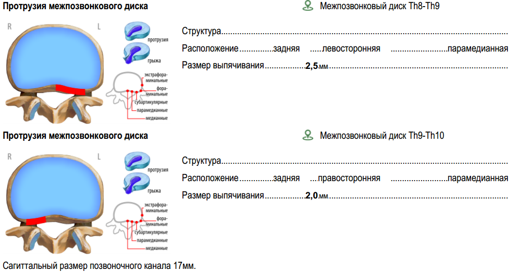
When single and multiple cysts are added to the study protocol, we get the following result:

A sedentary lifestyle, excess weight, curvature of the spine, weak back muscles, osteochondrosis, serious loads on the spine, and a metabolic disorder - this is what causes the appearance of changes in the intervertebral discs. The intervertebral disk receives all the nutrients from neighboring tissues through diffusion, since there are no blood vessels in the disk itself. It consists of the pulpal (gelatinous) nucleus and the fibrous ring surrounding it. When the metabolism stops, dystrophic changes begin in the fibrous ring, which leads to its thinning, loss of elasticity and the appearance of cracks, and the pulpous nucleus begins to slowly move to the side. When the gelatinous core reaches the edge of the disk, it begins to go beyond its limits, stretching the fibers of the annulus. So protrusion is formed. If the fibrous ring is torn,
For the description of protrusions and hernias complex localization is used. The pattern structure for them is the same:
<template><insertion-pictureid="3"is-short="true"selection-color="#FFB6AC" /><measurementid="2622"is-short="true"comment="Структура"/><lineis-short="true"><measurementid="8821"measurement-name="Расположение"comment="Расположение переднее\заднее"/><measurementid="8822"max-width="70"empty-name="true"comment="Расположение латеральное"/><measurementid="8823"empty-name="true"comment="Расположение сегментное"/></line><measurementid="2624"is-short="true"comment="Размер выпячивания диска"/><anatomy-commentcomment-id="279"comment-type="comment"spacing-before="NONE"/></template>Part of the MRI template of the thoracic spine:
<insertion-place><insertion-template-builderanatomy-width="145"name-width="28"status-width="30"localization-width="150"found-date-width="55"template-builder-width="45"anatomy-id="901"localization-id="869, 1501, 1502, 1503, 1504, 1505, 1506, 1507, 1508, 1509, 1510, 1511, 1450"id="582,542"use-template-builder-name="true"localization-marker-type="icon"/></insertion-place>In the id attribute, the value of 582 is the number of the protrusion pattern, and 542 is the hernia. Anatomy 901 ( anatomy-id attribute ) - herniated intervertebral disc.
Anatomy used for localization (localization-id)
- 869 - intervertebral disk C7-Th1;
- 1501 - intervertebral disk Th1-Th2;
- 1502 - intervertebral disk Th2-Th3;
- 1503 – межпозвонковый диск Тh3-Тh4;
- 1504 – межпозвонковый диск Тh4-Тh5;
- 1505 – межпозвонковый диск Тh5-Тh6;
- 1506 – межпозвонковый диск Тh6-Тh7;
- 1507 – межпозвонковый диск Тh7-Тh8;
- 1508 – межпозвонковый диск Тh8-Тh9;
- 1509 – межпозвонковый диск Тh9-Тh10;
- 1510 – межпозвонковый диск Тh10-Тh11;
- 1511 – межпозвонковый диск Тh11-Тh12;
- 1450 – межпозвонковый диск Th12-L1.


Additional anatomy
Consider the ultrasound pattern "soft tissues and lymph nodes of the neck, salivary glands", where you can describe the hyoid salivary gland.
Template sublingual salivary gland
<template><lineis-short="false"comment="Справа-Слева"><texttext-label=" " /><texttext-label="справа"max-width="193"is-color-selection="true"/><texttext-label="слева"max-width="193"is-color-selection="true"/></line><lineis-short="false"><measurementid="3969"comment="Структура подъязычных слюнных желез справа" /><measurementid="3981"max-width="193"empty-name="true"comment="слева" /></line><lineis-short="false"><measurementid="3970"comment="Контуры подъязычных слюнных желез справа" /><measurementid="3982"max-width="193"empty-name="true"comment="слева" /></line><lineis-short="false"><measurementid="3971"comment="Эхогенность подчелюстных слюнных желез справа" /><measurementid="3983"max-width="193"empty-name="true"comment="слева" /></line><lineis-short="false"><measurementid="3972"comment="Сосудистый рисунок подчелюстных слюнных желез справа" /><measurementid="3984"max-width="193"empty-name="true"comment="слева" /></line><lineis-short="false"><measurementid="3973"comment="Выводные протоки подъязычных слюнных желез справа" /><measurementid="3985"max-width="193"empty-name="true"comment="слева" /></line><lineis-short="false"><measurementid="12481"comment="Размер подъязычных слюнных желез справа" /><measurementid="12501"max-width="193"empty-name="true"comment="слева" /></line><anatomy-commentcomment-id="379"is-conclusion="false"spacing-before="NONE"comment="к подъязычным слюнным железам" /></template>Main pattern
<template><line><anatomyid="1261"font-size="10"font-bold="true"font-underline="false"is-short="false"comment="Мягкие ткани шеи" /><measurementid="3861"comment="Структура мягких тканей шеи" /></line><lineis-short="false"comment="Справа-Слева"><texttext-label=" " /><texttext-label="справа"max-width="193"is-color-selection="true"/><texttext-label="слева"max-width="193"is-color-selection="true"/></line><template-builderid="597"comment="Подчелюстные слюнные железы"/><template-builderid="598"comment="Околоушные слюнные железы"/><insertion-place><insertion-template-builderanatomy-width="180"need-template-builder="false"anatomy-id="1283"id="599"comment="Подъязычные слюнные железы"/><insertion-template-builderanatomy-width="180"need-template-builder="false"anatomy-id="1307"id="595 comment="Лимфатические узлы"/></insertion-place><anatomy-commentcomment-id="111"/><conclusion-labelspacing-before="HALF"/><anatomy-commentcomment-id="51"comment-type="conclusion"/></template>

Additional measurements
The previously considered Bauer reflex is in the insert with the reflexes of children up to one year old.
Insert Template:
<template><line><measurementid="8621"comment="Сосательный рефлекс" /><measurementid="8622"comment="Поисковый рефлекс" /></line><line><measurementid="8623"comment="Хоботковый рефлекс" /><measurementid="8624"comment="Ладонно-ротовой рефлекс" /></line><line><measurementid="8625"comment="Рефлекс Моро" /><measurementid="8626"comment="Хватательный рефлекс" /></line><line><measurementid="8627"comment="Защитный рефлекс" /><measurementid="8628"comment="Рефлекс Галанта" /></line><line><measurementid="8629"comment="Рефлекс Бауэра" /><measurementid="8630"comment="Опорно-шаговый рефлекс" /></line></template>Part of the pediatrician examination template:
<insertion-place><!-- Измерения для детей до года --><insertion-template-builderfont-bold="false"font-underline="true"need-template-builder="false"name-overriden-button="Измерения для детей до года"name-overriden="Измерения для детей до года"anatomy-width="200"id="921"/><!-- Рефлексы детей до года --><insertion-template-builderfont-bold="false"font-underline="true"need-template-builder="false"name-overriden-button="Рефлексы детей до года"name-overriden="Рефлексы детей до года"anatomy-width="200"id="922"/></insertion-place>

We will deal with the addition of a confirmatory test for hepatitis C. The test pattern itself:
<template><measurementid="2365"comment="Подтверждающий тест на антитела к вирусу гепатита С (Anti HCV)"/><measurementid="2362"comment="Антитела к структурному белку Core вируса гепатита С (Anti HCV-Core)"/><measurementid="2381"comment="Антитела к неструктурному белку NS3 вируса гепатита С (Anti HCV-NS3)"/><measurementid="2363"comment="Антитела к неструктурному белку NS4 вируса гепатита С (Anti HCV-NS4)"/><measurementid="2364"comment="Антитела к неструктурному белку NS5 вируса гепатита С (Anti HCV-NS5)"/></template>Template "Anti-HCV Antibodies (Anti HCV)":
<template><measurementid="90"comment="Гепатит С (Anti HCV) IgG"/><insertion-place><insertion-template-builderfont-bold="false"font-underline="true"need-template-builder="false"anatomy-width="180"name-overriden="Гепатит С. подтверждающий тест"id="572"/></insertion-place><anatomy-commentcomment-id="355"comment-type="comment"spacing-before="NONE"comment="Кровь. Антитела к Гепатиту С"/></template>

Description of the removed organs
As you know, a person can adapt to life without certain organs. However, history remembers times when wild thoughts about “extra organs” appeared in the minds of scientists, which do not affect the vital activity of the organism and are subject to removal, and the sad consequences of their realization in reality. So in the late XIX - early XX century, this idea reached its apogee.
More about dashing times
Чарльз Дарвин в своей книге «Происхождение человека и половой отбор» (1871 г.) перечислил ряд рудиментарных органов (аппендикс, копчиковая кость, зубы мудрости, мышцы уха, волосы на теле и т. д.), наличие которых, по его мнению, подтверждало теорию об эволюции.
Немецкий анатом Роберт Видерсгейм в работе «Строение человека со сравнительно-анатомической точки зрения» (1890 г.) дал обширный список рудиментарных органов. К ранее рассмотренным Дарвином добавились миндалины, щитовидная железа, слёзные железы, некоторые клапаны вен и т. д.
Русский биолог Илья Ильич Мечников в своем труде «Этюды о природе человека» (1913 г.) упоминает работы Дарвина и Видерсгейма, а также рассуждает о несовершенстве и дисгармонии человеческого организма. Одна из глав посвящена пищеварительной системе, где упоминается бесполезность аппендикса и всячески отвергается предположение, высказанное голландским врачом Кольбругге, о полезности червеобразного отростка (аппендикса), так как там развивается кишечная бактерия, предохраняющая организм от некоторых болезней пищеварительной системы. Однако никаких весомых доказательств Кольбругге в пользу своей теории не предъявлял. В опалу Мечникова попала и толстая кишка, полезные функции которой сводились к минимуму, а вот проблемы, связанные с ней, наоборот, весьма подробно рассматривались (запоры, всасывание вредных веществ, очаг опасных болезней и частое место злокачественных опухолей). Поэтому, на взгляд Мечникова, удаление этого органа в целом привело бы к положительным результатам. Хоть сам ученый и не проверял свое утверждение, но оно нашло подтверждение в практике некоторых хирургов, проводивших подобные операции. Были и врачи, которые с большим энтузиазмом отнеслись к его словам и отрезали орган порой без всякой необходимости. Однако не все пациенты выживали или могли продолжить свою обычную жизнь. Только через десятилетия подобный метод лечения подвергся серьезной критике.
Идея заблаговременного удаления аппендикса многим врачам пришлась по вкусу, так как от аппендицита умирало немало пациентов. Не стеснялись удалять и миндалины с гландами. Наибольшее распространение идея удаления этих органов у детей в раннем возрасте получила в Европе и США. Однако спустя некоторое время специалистами были осознаны их ошибки. Дети без аппендикса плохо усваивали материнское молоко, отставали в развитии, чаще болели и испытывали проблемы с пищеварением. Врачам было не понятно, почему так происходило. Только в 2007 году американские ученые из университета Дьюка (Duke University) выяснили, что в аппендиксе находятся бактерии необходимые для нормальной работы кишечника. Роль миндалин и гланд узнали также позже, а до того момента врачи заметили, что дети без них чаще остальных подвержены болезням бронхов и легких.
В целом развитие идеи «лишних» органов можно объяснить банальным незнанием о работе и функциях предполагаемых рудиментов.
Порой практиковались достаточно странные методы лечения некоторых заболеваний. Некоторые из них основывались на удалении органов. Например:
- лечение женской истерии, начавшееся в викторианскую эпоху и закончившееся в Европе в конце XIX века, а в Америке – в начале XX века. Удалению подлежали женские половые органы;
- лечение психических заболеваний Генри Коттоном. Врач считал, что такого рода заболевания вызывают инфекции. Сначала он удалял пациентам зубы. Если эффекта не было, то в ход шли остальные органы (гланды, желчный пузырь и т. д.).
Today, doctors use the removal of organs and anatomical parts of the body (ectomy) only in extreme cases: injuries or serious pathologies. It can be removed as one of the paired organs (eyes, kidney, lung, etc.) in the case of normal operation of the other, or unpaired. The latter include:
- stomach - in the treatment of gastric cancer. After gastrectomy, the esophagus and small intestine are connected;
- spleen - in case of blood diseases, injuries and other pathological conditions;
- gallbladder - with cholecystitis;
- part of the liver - can be removed up to 75%. The liver has a regenerative function, which occurs very slowly;
- pancreas;
- colon - with Crohn's disease and malignant tumors;
- thyroid;
- etc.
An anatomy-status tag has been added to capture information about a remote organ or anatomical part of the body .
Anatomy-status
It is a drop-down list of three statuses: yes, no, not visualized. To correctly display the study protocol, it is necessary to take into account the peculiarities of the language. So for each gender and number, you can adjust the status options separately, and for anatomy - the desired list. For example:
- feminine, single number - is (visualized as a space in the document), deleted, not visualized;
- masculine unit number - “space”, deleted, not rendered;
- the plural is “space”, deleted, not rendered.
In addition to the obvious statuses, there is / no "not rendered" was added. As the name implies, it is used in cases where a body is not visible during the examination. For example, due to the accumulation of gas in the intestine, some abdominal organs may not be visible.
In general, an entity with anatomy statuses is used not only for the anatomy-status tag and organs to be removed, but also in inserts with the type “education”. For example:
- for the fetus - visualized, born, not visualized;
- for formations (cyst, node, etc.) - it is visualized, promptly removed, not visualized.
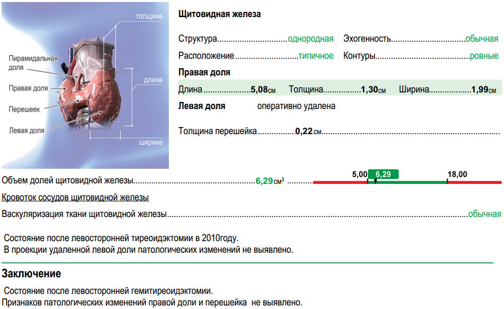
The anatomy-status tag works as follows: if you select the “no” and “not visualized” statuses, all dimensions related to this anatomy are removed, and a line with the name of the anatomy and its status remains. In order for this to work, all the code related to the anatomy to be removed is separated into a separate template and a link to it ( template-builder tag ) in the main template is written.
When the status of anatomy is set to "no" or "not visualized," it is stored in anatomy-addon .
Thus, the following anatomy can fall into the essence of anatomy-addon :
- changed anatomy, which usually do not describe;
- remote anatomy;
- appeared "non-standard" anatomy.
The anatomy-status tag contains several attributes:
- anatomy-id - the number of anatomy in the database;
- max-width - the overall width of the element;
- spacing-before, is-short .
The thyroid ultrasound pattern is one of those patterns that have undergone significant structural changes during system design.
The very first version of the thyroid ultrasound template
<templateimage-id="5"need-warning="true"image-position="left-top-corner"><anatomyid="22"font-size="10"font-bold="true"font-underline="false"is-short="true"comment="Щитовидная железа" /><lineis-short="true"spacing-before="HALF"><measurementid="310"max-width="156"comment="Эхоструктура"/><measurementid="341"max-width="156"comment="Эхогенность"/></line><lineis-short="true"><measurementid="308"max-width="156"comment="Расположение"/><measurementid="342"max-width="156"comment="Контуры"/></line><anatomyid="24"font-size="10"font-bold="false"font-underline="false"anatomy-name="Размеры правой доли щитовидной железы:"is-short="true"spacing-before="HALF"/><measurement-groupid="2"is-short="true"is-color-selection="true"/><anatomyid="23"font-size="10"font-bold="false"font-underline="false"anatomy-name="Размеры левой доли щитовидной железы:"is-short="true"spacing-before="HALF"/><measurement-groupid="1"is-short="true"is-color-selection="true"/><measurementid="307"is-short="true"measurement-name="Толщина перешейка"spacing-before="HALF"/><measurementid="309"measurement-name="Объем долей щитовидной железы"/><anatomy-commentcomment-id="9"is-conclusion="false"spacing-before="HALF"/><anatomy-commentcomment-id="8"is-conclusion="true"spacing-before="HALF"/></template>Adding template-builder and anatomy-status tags and inserts
Основной шаблон:
Правая доля:
Левая доля:
<templateimage-id="5"need-warning="true"image-position="left-top-corner"><anatomyid="22"font-size="10"font-bold="true"font-underline="false"is-short="true"comment="Щитовидная железа"/><lineis-short="true"spacing-before="HALF"><measurementid="310"max-width="156"comment="Эхоструктура"/><measurementid="341"max-width="156"comment="Эхогенность"/></line><lineis-short="true"><measurementid="308"max-width="156"comment="Расположение"/><measurementid="342"max-width="156"comment="Контуры"/></line><template-builderid="253"is-short="true"/><template-builderid="254"is-short="true"/><measurementid="307"is-short="true"measurement-name="Толщина перешейка"spacing-before="HALF" /><measurementid="309"measurement-name="Объем долей щитовидной железы"/><insertion-place><!-- узел --><insertion-template-builderanatomy-id="1041"anatomy-width="100"name-width="136"need-template-builder="false"localization-id="23,1021,1022,1023,24,1024,1025,1026,25"need-status="false"localization-width="177"localization-marker-type="word"id="578"/><insertion-template-builderanatomy-id="1042"anatomy-width="100"name-width="136"need-template-builder="false"localization-id="23,1021,1022,1023,24,1024,1025,1026,25"need-status="false"localization-width="177"localization-marker-type="word"id="580"/><!-- киста --><insertion-template-builderanatomy-id="821"anatomy-width="100"name-width="136"need-template-builder="false"localization-id="23,1021,1022,1023,24,1024,1025,1026,25"need-status="false"localization-width="177"localization-marker-type="word"id="538"/><insertion-template-builderanatomy-id="822"anatomy-width="100"name-width="136"need-template-builder="false"localization-id="23,1021,1022,1023,24,1024,1025,1026,25"need-status="false"localization-width="177"localization-marker-type="word"id="540"/><!-- Кровоток --><insertion-template-builderfont-bold="false"font-underline="true"need-template-builder="false"anatomy-width="180"name-overriden="Кровоток сосудов щитовидной железы"id="537"/></insertion-place><anatomy-commentcomment-id="9"comment-type="comment"spacing-before="HALF"/><conclusion-labelspacing-before="HALF"/><anatomy-commentcomment-id="8"comment-type="conclusion"/></template>Правая доля:
<template><lineis-short="true"><anatomyid="24"font-size="10"font-bold="true"font-underline="false"spacing-before="HALF"comment="Правая доля"/><anatomy-statusanatomy-id="24" /></line><measurement-groupid="2"is-color-selection="true"is-short="true"/></template>Левая доля:
<template><lineis-short="true"><anatomyid="23"font-size="10"font-bold="true"font-underline="false"spacing-before="HALF"comment="Левая доля"/><anatomy-statusanatomy-id="23" /></line><measurement-groupid="1"is-color-selection="true"is-short="true"/></template>
When we started, each research protocol template consisted of a comment and a conclusion. The desire for formalization resulted primarily in the creation of a system for storing numerical data and a description of human anatomy. As a result, comments declined and were used as explanations of the main data.
If you look at the conclusions, then among the information that usually gets there, we can distinguish the following types of data:
- diagnoses:
- "Signs of chronic tonsillitis"
- "duodenitis?"
- "K21.0 GERD with esophagitis"
- destination:
- "Recommended diet with restriction of fat"
- "Chlorhexidine 2-3 jets 2 times a day"
- directions:
- "general urine analysis"
- "MRI of the cervical spine"
- "Consultation endocrinologist"
Thus, the conclusion remains the "den" of non-formalized information. Therefore, the further development of the medical data storage system is aimed at formalizing these entities.
