New Type SERS Sensor Helps in Early Cancer Detection
Raman spectroscopy is a technology that allows you to identify cancer cells among a large number of healthy ones, distinguish fake pictures from real ones and conduct a large number of other studies.
Nevertheless, the widespread adoption of such technology is hindered by the high cost of electronic components from which such systems are created. However, the technology may still be widely used in everyday life, and not just in laboratories, thanks to the new achievement of an integrated team of specialists led by scientists from the University of Buffalo.
Scientists have created miniature and inexpensive SERS sensors that can be used, including in mobile devices.
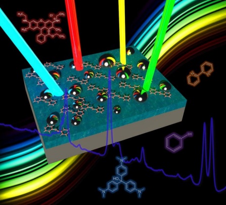
SERS technology is based on the analysis of variable-wavelength laser beams reflected from the material being studied. The achievement is to create a substrate material on which the samples are placed. This substrate is universal, and in a certain way allows you to enhance the "reverse reaction" of the molecules of the compounds that are treated with a laser.
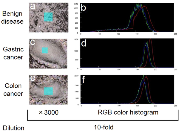
Volumetric and color histograms of the surface structure of silver NHC chips with tissue samples using a 3D laser scanning confocal microscope
The essence of the method is that a beam with a certain wavelength is passed through a sample of the test substance, which scatters upon contact with the sample. The rays obtained are collected using a lens into a single beam and passed through a filter that separates weak (0.001% intensity) Raman rays from more intense (99.999%) Rayleigh rays. "Pure" Raman rays are amplified and sent to a detector, which records the frequency of their oscillations.
The positive side of the method is that you can use ultra-small amounts of the substance. Experts say that you can even work with samples that are not visible to the naked eye. In this case, the substrate improves the light field, which enhances the "inelastic scattering." Scientists from the University of Buffalo were able to create a universal substrate for this kind of research, while the usual Raman spectroscopy requires the use of different substrates to study various materials. In addition, previously it was required to use lasers with different wavelengths and different powers.
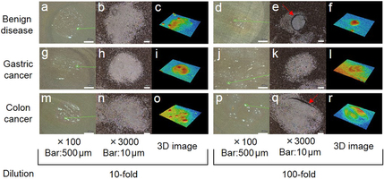
The results of the analysis of tissues of various types (including cancers)
The universal substrate, created by specialists from the University of Buffalo and Fudan University, is a nanostructured broadband network. This network can pick up light radiation with different wavelengths (from 450 to 1100 nm), which is used by SERS. The substrate consists of several layers, one of which is a mirror layer of aluminum or silver. The dielectric is a layer of silicon or aluminum oxide. The dielectric separates the “mirror” and silver nanoparticles.
“All this acts like a master key. Instead of using different substrates for light radiation with different wavelengths, we use only one substrate. It's kind of a master key that opens up a lot of doors, ”says Nan Zhang, a team of scientists.
Zang also claims that the use of such a chip is possible in various fields of science and industry. “The ability to work with even a very small amount of the substance can help determine cancer, malaria, HIV and other work,” Nan Zang comments.
As mentioned above, the new development can be used not only in medicine. For example, this technology will help to distinguish the original picture from a fake, to determine the concentration of toxins in air or water, to determine the constituent components of a chemical weapon during its use.
Now scientists from the project group are engaged in various studies in the field of nanophotonics, biophotonics, the study of hybrid materials and their derivatives, nonlinear and fiber optics, metamaterials, optofluids and much more. Thanks to the work of specialists studying related industries, it became possible to create a universal SERS chip.