Scientists have developed an atomic force microscope that allows you to observe the dynamics of neurons at the nano level
Researchers at the Max Planck Florida Institute for Neuroscience and Kanazawa University (Japan) were able to obtain images of the structural dynamics of living neurons with unprecedented spatial resolution. The

researchers modernized the atomic force microscope they have at their disposal. AFM). Thanks to this modernization, he became able to record dynamic changes in the structure of living neurons with spatial resolution and speed unprecedented at the present time.
- Atomic force microscopy (AFM) is a leading tool for working with images, measuring and manipulating materials with atomic resolution - on the order of fractions of a nanometer.
- AFM receives images of the surface topography of the structure literally “touching” the surface by scanning with a very thin needle (tip diameter of about 5 nm, 1/100 of the light wavelength or 1/10000 of the hair)
- This technology is used to image solid materials. But it was difficult to use AFM for soft and large samples such as eukaryotic cells and neurons without damaging the sample. In addition, conventional AFM imaging is too slow to capture rapid changes in cell morphology.
- This new system allows you to analyze morphological changes ~ 20-100 times faster than a standard light microscope
Scientists, developing technology for high-speed imaging (long-tip high-speed, LT-HS-AFM) have found a way to avoid damage to biological samples. They used an extremely long and sharp probe mounted on a flexible plate, such a soft and pliable console of the new microscope provides minimal probe pressure on the sample. A high-speed optical system that operates at a frequency of 800 kHz (thousands of times per second) controls the movement of the probe tip.
The new microscope, which does not damage biological tissues, is optimized for high-speed imaging; it takes only a few seconds to create a single frame. This allows you to register quite dynamic processes that occur in living cells, while obtaining spatial resolution that is hundreds of times higher than the capabilities of the best optical microscopes.
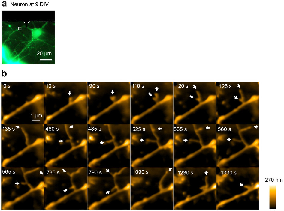
Image of a hippocampal neuron transfected with mEGFP. White dashed lines show the shadow of the cantilever. The white square shows the image area of the LT-HS-AFM.
With the help of the device they created, scientists were able to get a series of images that made it possible to track the dynamics of structural changes in the surface of cells, the so-called process of morphogenesis, the processes of membrane ripple formation, the formation of depressions and other changes caused by forced stimulation of various types.
researchers modernized the atomic force microscope they have at their disposal. AFM). Thanks to this modernization, he became able to record dynamic changes in the structure of living neurons with spatial resolution and speed unprecedented at the present time.
- Atomic force microscopy (AFM) is a leading tool for working with images, measuring and manipulating materials with atomic resolution - on the order of fractions of a nanometer.
- AFM receives images of the surface topography of the structure literally “touching” the surface by scanning with a very thin needle (tip diameter of about 5 nm, 1/100 of the light wavelength or 1/10000 of the hair)
- This technology is used to image solid materials. But it was difficult to use AFM for soft and large samples such as eukaryotic cells and neurons without damaging the sample. In addition, conventional AFM imaging is too slow to capture rapid changes in cell morphology.
- This new system allows you to analyze morphological changes ~ 20-100 times faster than a standard light microscope
Scientists, developing technology for high-speed imaging (long-tip high-speed, LT-HS-AFM) have found a way to avoid damage to biological samples. They used an extremely long and sharp probe mounted on a flexible plate, such a soft and pliable console of the new microscope provides minimal probe pressure on the sample. A high-speed optical system that operates at a frequency of 800 kHz (thousands of times per second) controls the movement of the probe tip.
Process video
The new microscope, which does not damage biological tissues, is optimized for high-speed imaging; it takes only a few seconds to create a single frame. This allows you to register quite dynamic processes that occur in living cells, while obtaining spatial resolution that is hundreds of times higher than the capabilities of the best optical microscopes.
Image of a hippocampal neuron transfected with mEGFP. White dashed lines show the shadow of the cantilever. The white square shows the image area of the LT-HS-AFM.
540nm
710nm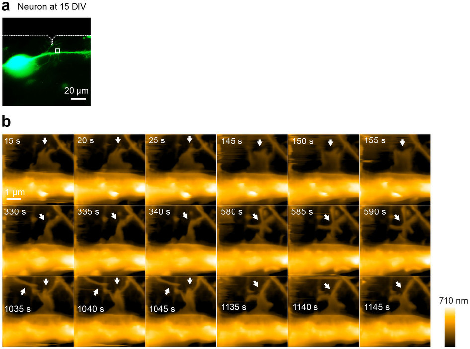
With the help of the device they created, scientists were able to get a series of images that made it possible to track the dynamics of structural changes in the surface of cells, the so-called process of morphogenesis, the processes of membrane ripple formation, the formation of depressions and other changes caused by forced stimulation of various types.
In the very near future, we plan on the basis of existing technology to create a technology for visualizing the synapse morphology in real time, while obtaining a sub-nanometer resolution, "says Ryohei Yasuda, a scientist from the Max Planck Institute, -" These data have it is very important for modern science, because changes in the morphology of synapses are the basis of synaptic plasticity, the basis of our memory and cognitive function. Investigating these processes, we can learn a lot about neurons to store information, and this will ultimately help us to come close to being able to generate artificial memories, instant training, and many other things that are so far only fiction